

Modalities for Visualization of Cortical Bone Remodeling: The Past, Present, and Future
- PMID: 26322017
- PMCID: PMC4531299
- DOI: 10.3389/fendo.2015.00122
Modalities for Visualization of Cortical Bone Remodeling: The Past, Present, and Future
Abstract
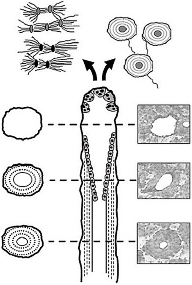
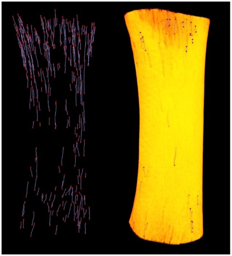
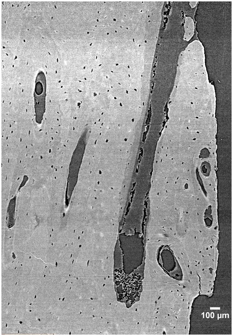
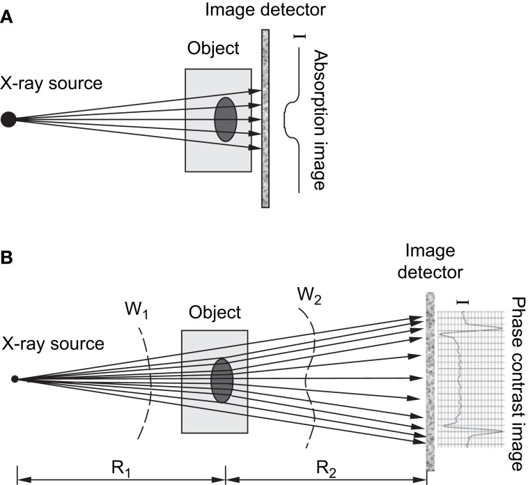
Bone's ability to respond to load-related phenomena and repair microdamage is achieved through the remodeling process, which renews bone by activating groups of cells known as basic multicellular units (BMUs). The products of BMUs, secondary osteons, have been extensively studied via classic two-dimensional techniques, which have provided a wealth of information on how histomorphology relates to skeletal structure and function. Remodeling is critical in maintaining healthy bone tissue; however, in osteoporotic bone, imbalanced resorption results in increased bone fragility and fracture. With increasing life expectancy, such degenerative bone diseases are a growing concern. The three-dimensional (3D) morphology of BMUs and their correlation to function, however, are not well-characterized and little is known about the specific mechanisms that initiate and regulate their activity within cortical bone. We believe a key limitation has been the lack of 3D information about BMU morphology and activity. Thus, this paper reviews methodologies for 3D investigation of cortical bone remodeling and, specifically, structures associated with BMU activity (resorption spaces) and the structures they create (secondary osteons), spanning from histology to modern ex vivo imaging modalities, culminating with the growing potential of in vivo imaging. This collection of papers focuses on the theme of "putting the 'why' back into bone architecture." Remodeling is one of two mechanisms "how" bone structure is dynamically modified and thus an improved 3D understanding of this fundamental process is crucial to ultimately understanding the "why."
Keywords: basic multicellular unit; bone; micro-CT; remodeling; synchrotron.
Figures
References
-
- Havers C. Osteologia Nova, or, Some New Observations of the Bones and the Parts Belonging to them, with the Manner of their Accretion, and Nutrition, Communicated to the Royal Society in Several Discourses. Ann Arbor, MI: University Microfilms International; (1691).
-
- Tomes J, Morgan CD. Observations on the structure and development of bone. Philos Trans R Soc Lond B Biol Sci (1853) 143:109–39.10.1098/rstl.1853.0004 - DOI
-
- Wolff J. Das Gesetz der Transformation der Knochen. Berlin: Hirschwald Verlag; (1892).
-
- Johnson L. Morphologic analysis of pathology. In: Frost H, editor. Bone Biodynamics. Boston, MA: Little, Brown, and Company; (1964), 543–54.
Publication types
LinkOut - more resources
Full Text Sources
Other Literature Sources
Miscellaneous