Review
. 2015 Aug 12:6:417.
doi: 10.3389/fimmu.2015.00417. eCollection 2015.

Complement in the Homeostatic and Ischemic Brain

Affiliations
Review

Complement in the Homeostatic and Ischemic Brain

Ali Alawieh et al. Front Immunol. .

Abstract

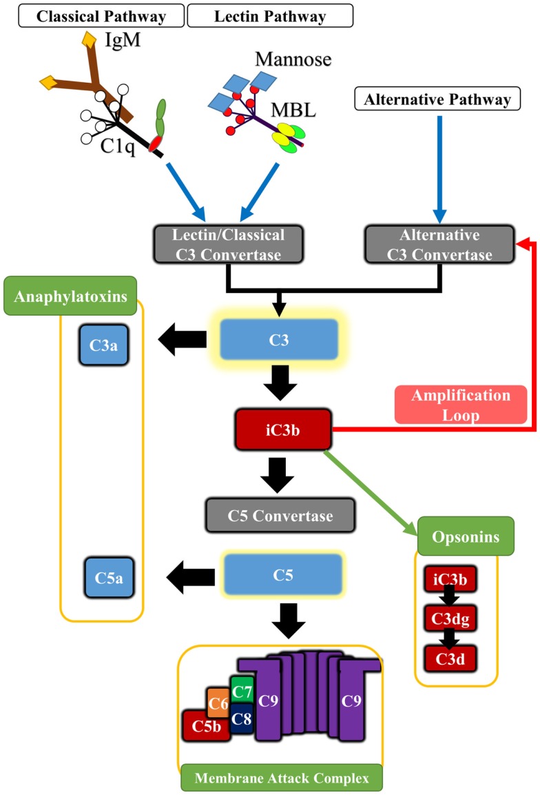
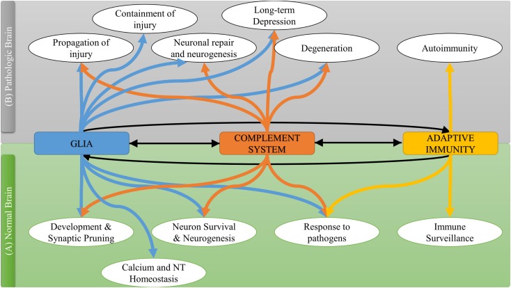
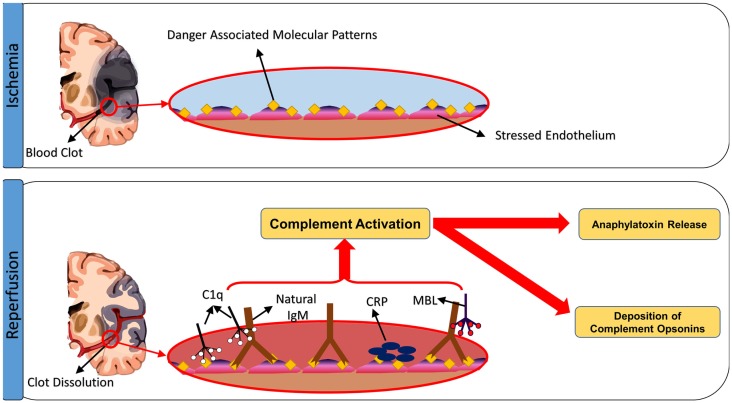
The complement system is a component of the immune system involved in both recognition and response to pathogens, and it is implicated in an increasing number of homeostatic and disease processes. It is well documented that reperfusion of ischemic tissue results in complement activation and an inflammatory response that causes post-reperfusion injury. This occurs following cerebral ischemia and reperfusion and triggers secondary damage that extends beyond the initial infarcted area, an outcome that has rationalized the use of complement inhibitors as candidate therapeutics after stroke. In the central nervous system, however, recent studies have revealed that complement also has essential roles in synaptic pruning, neurogenesis, and neuronal migration. In the context of recovery after stroke, these apparent divergent functions of complement may account for findings that the protective effect of complement inhibition in the acute phase after stroke is not always maintained in the subacute and chronic phases. The development of effective stroke therapies based on modulation of the complement system will require a detailed understanding of complement-dependent processes in both early neurodegenerative events and delayed neuro-reparatory processes. Here, we review the role of complement in normal brain physiology, the events initiating complement activation after cerebral ischemia-reperfusion injury, and the contribution of complement to both injury and recovery. We also discuss how the design of future experiments may better characterize the dual role of complement in recovery after ischemic stroke.

Keywords: brain ischemia; complement; innate immunity; neuroprotection; reperfusion injury; stroke.


Figures

Figure 1
Figure 1
Basic outline of the principle pathways of complement activation and complement effector molecules. The classical pathway is triggered by the binding of C1q to antibody Fc regions, pentraxins or certain cell surface determinants. The lectin pathway is triggered by Mannose-binding lectin (MBL) or ficolins which bind to carbohydrate patterns, including those found on some IgM antibodies. Both pathways lead to cleavage of C2 and C4 components, forming the classical pathway C3 convertase (C4b2a) that cleaves C3. The alternative pathway is spontaneously activated forming the alternative pathway C3 convertase (C3bBb), and it also serves to amplify classical and lectin pathway activation. Cleavage of C3 leads to the generation of C3a and C3b. Activated C3b deposits on cell surfaces and is further degraded to iC3b, C3dg, and C3d, which serve as opsonins for receptors on immune cells. In addition, C3b associates with preformed C3 convertase, forming C5 convertase that in turn cleaves C5 into C5a and C5b. Deposition of C5b on cell surface initiates assembly of the cytolytic membrane attack complex (MAC or C5b-9). The C5a and C3a anaphylatoxins are potent pro-inflammatory molecules, and also modulate various homeostatic effects through G-protein signaling.
Figure 2
Figure 2
The interplay between complement system and other immune components in normal and pathological brain.
Figure 3
Figure 3
Triggers of complement activation after cerebral ischemia-reperfusion injury. Ischemic insult induces expression of neoepitopes or danger-associated molecular patterns (DAMPs) on the surface of stressed endothelial cells. The exposed DAMPs are recognized by circulating natural self-reactive antibodies, principally IgM, which triggers complement activation. Although IgM binds C1q, it appears to be the binding of MBL and activation of the lectin pathway that drives ischemia and reperfusion injury in the organs systems examined, including the brain. Complement can be also activated through direct binding of C1q to apoptotic cells, as well as through C-reactive protein-induced complement activation.

References

    1. Barber PA, Davis SM, Infeld B, Baird AE, Donnan GA, Jolley D, et al. Spontaneous reperfusion after ischemic stroke is associated with improved outcome. Stroke (1998) 29:2522–8.10.1161/01.STR.29.12.2522 - DOI - PubMed
    1. Patel RD, Saver JL. Evolution of reperfusion therapies for acute brain and acute myocardial ischemia: a systematic, comparative analysis. Stroke (2013) 44:94–8.10.1161/STROKEAHA.112.666925 - DOI - PubMed
    1. Iadecola C, Anrather J. The immunology of stroke: from mechanisms to translation. Nat Med (2011) 17:796–808.10.1038/nm.2399 - DOI - PMC - PubMed
    1. Barber PA, Zhang J, Demchuk AM, Hill MD, Buchan AM. Why are stroke patients excluded from TPA therapy? An analysis of patient eligibility. Neurology (2001) 56:1015–20.10.1212/WNL.56.8.1015 - DOI - PubMed
    1. Katzan IL, Hammer MD, Hixson ED, Furlan AJ, Abou-Chebl A, Nadzam DM, et al. Utilization of intravenous tissue plasminogen activator for acute ischemic stroke. Arch Neurol (2004) 61:346–50.10.1001/archneur.61.3.346 - DOI - PubMed