Review
. 2015 Aug 26;6(3):162-208.
doi: 10.4331/wjbc.v6.i3.162.

Roles of the canonical myomiRs miR-1, -133 and -206 in cell development and disease

Affiliations
Review

Roles of the canonical myomiRs miR-1, -133 and -206 in cell development and disease

Keith Richard Mitchelson et al. World J Biol Chem. .

Abstract

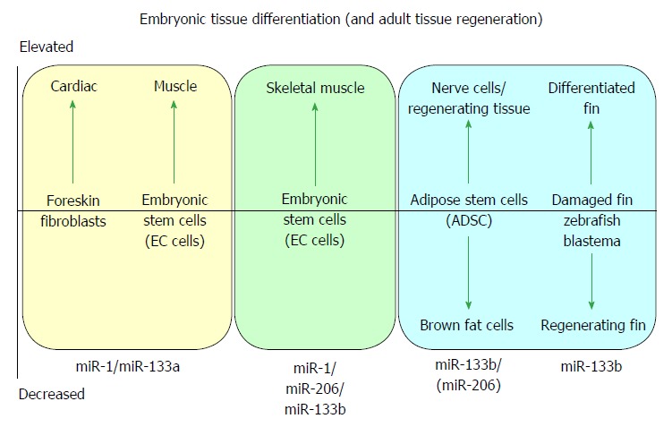
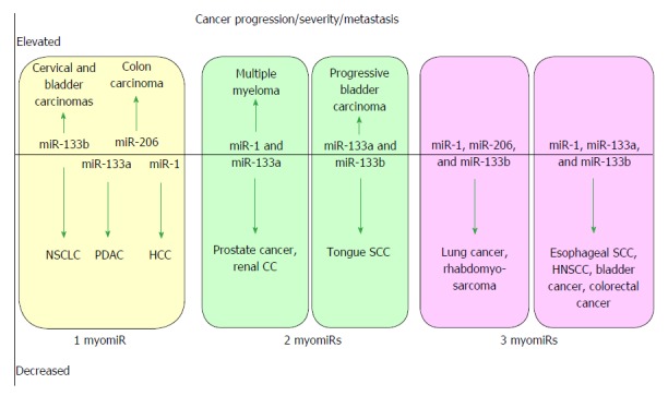
MicroRNAs are small non-coding RNAs that participate in different biological processes, providing subtle combinational regulation of cellular pathways, often by regulating components of signalling pathways. Aberrant expression of miRNAs is an important factor in the development and progression of disease. The canonical myomiRs (miR-1, -133 and -206) are central to the development and health of mammalian skeletal and cardiac muscles, but new findings show they have regulatory roles in the development of other mammalian non-muscle tissues, including nerve, brain structures, adipose and some specialised immunological cells. Moreover, the deregulation of myomiR expression is associated with a variety of different cancers, where typically they have tumor suppressor functions, although examples of an oncogenic role illustrate their diverse function in different cell environments. This review examines the involvement of the related myomiRs at the crossroads between cell development/tissue regeneration/tissue inflammation responses, and cancer development.

Keywords: Cancer; Cell development; MiR-1; MiR-133a; MiR-133b; MiR-206; Muscle microRNAs.


Figures

Figure 1
Figure 1
The roles of the myomiRs during embryonic tissue differentiation and adult tissue regeneration. Elevated levels of miR-1 and miR-133a are essential for differentiation of cardiac muscle[10,15], whilst miR-1, miR-206 and miR-133b are required for skeletal muscle differentiation[7,13]. Elevation of miR-133b levels in adipose stem cells leads to differentiation to a nerve-cell like fate[71], whilst reduction in miR-133b leads to brown fat cell differentiation[74]. Strong depletion of miR-133b and elevated Fgf allows regeneration of damaged zebrafish appendages[67,68].
Figure 2
Figure 2
Alignment of the principal transcripts of the miR-206/miR-133b locus of (A) mouse chromosome 1 and (B) human chromosome 9, compiled from several sources. A: The primary long intergenic non-coding polyadenylated RNA (linc-MD1) of 15 kb is initiated at the upstream distal promoter (DIST) and encompasses both microRNA-206 and -133b genes, however maturation of the pri-linc-MD1 results only in the release of pre-miR-133b which maturates into mir-133b which accumulates in the cell nucleus, as well as the mature 521 bp spliced linc-MD1 RNA which accumulates in the cytoplasm of skeletal muscle cells[38]. The mature linc-MD1 RNA sequence contains binding sites for miR-135 and miR-133 and acts as a complementary “sponge” to regulate their abundance and regulate expression of their target genes including key muscle transcription factors. Accumulation of linc-MD1 RNA and miR-133b are mutually exclusive. The DIST promoter contains E-box sequences recognized by MyoD and is active in differentiating muscle cells when increasing levels of MyoD induce the expression of linc-MD1 or miR-133b. In contrast, the primary transcript of miR-206 initiates at an independent proximal promoter (PROX) which is located intronic to linc-MD1 gene and is already active in undifferentiated proliferating cells. The PROX promoter binds both MyoD and myogenin (ChiP data)[38] and is functional in the presence of myogenin alone, although increased expression activity is also associated with differentiation of muscle cells[11]. The primary expressed transcript for miR-206 is likely coincident with the 5.3 kb random cloned mouse transcript cDNA, GenBank sequence AK132542[40], although the first 13 nt of the pre-mir-206 sequence is absent from that sequence. A second polyadenylated non coding RNA 7H4 was previously identified in rat muscle cells which are specifically associated with nerve synapses[39] and is expressed at rat development stages coincident with the strong expression of MyoD and myogenin[11]. The 5.2 kb long 7H4 ncRNA clone is slightly truncated at the 5’ terminus compared to the AK132542 sequence, and may represent a processed product from which pre-miR-206 has been cleaved[8]. A second 1.6 kb short 7H4 ncRNA transcript, exactly coincident with the 3’ terminal region of the longer transcript, is also abundant in rat muscle[39] and may represent a second processing product; B: The lnc RNA RP11-771D21.2 (Hs.582788) has been identified in RNA seq libraries and aligned with known genomic loci. Information about the regulation of its expression is not available.
Figure 3
Figure 3
Roles of the myomiRs during cancer progression. Altered expression of myomiRs is associated with cancer progression and metastasis. Note that the changed level of myomiR typically contributes to cancer progression. Reduced levels of a single myomiR are associated with PDAC[103], NSCLC[105] and HCC[98], whilst an elevated single miR is associated with cervical carcinoma[121] and bladder cancer (TCC)[122]. Increased expression of miR-1/-133a is associated with t(14;16) translocation multiple myeloma[152], whilst reduced expression is associated with prostate cancer[102] and RCC[138], respectively. Reduced expression of other myomiR combinations is also associated with TSCC[111], ESCC[132], HNSCC[114,143], GIST[281], bladder cancer (TCC)[93], lung cancer[85,99], colorectal cancer[86,87] and rhabdomyosarcoma[88].

Similar articles

Cited by

References

    1. McCarthy JJ. The MyomiR network in skeletal muscle plasticity. Exerc Sport Sci Rev. 2011;39:150–154. - PMC - PubMed
    1. Liu N, Olson EN. MicroRNA regulatory networks in cardiovascular development. Dev Cell. 2010;18:510–525. - PMC - PubMed
    1. Bassel-Duby R, Olson EN. Signaling pathways in skeletal muscle remodeling. Annu Rev Biochem. 2006;75:19–37. - PubMed
    1. Potthoff MJ, Olson EN, Bassel-Duby R. Skeletal muscle remodeling. Curr Opin Rheumatol. 2007;19:542–549. - PubMed
    1. Townley-Tilson WH, Callis TE, Wang D. MicroRNAs 1, 133, and 206: critical factors of skeletal and cardiac muscle development, function, and disease. Int J Biochem Cell Biol. 2010;42:1252–1255. - PMC - PubMed

LinkOut - more resources