

Adipose tissue-derived stem cells as a therapeutic tool for cardiovascular disease
- PMID: 26322185
- PMCID: PMC4549779
- DOI: 10.4330/wjc.v7.i8.454
Adipose tissue-derived stem cells as a therapeutic tool for cardiovascular disease
Abstract
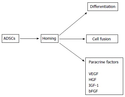
Adipose tissue-derived stem cells (ADSCs) are adult stem cells that can be easily harvested from subcutaneous adipose tissue. Many studies have demonstrated that ADSCs differentiate into vascular endothelial cells (VECs), vascular smooth muscle cells (VSMCs), and cardiomyocytes in vitro and in vivo. However, ADSCs may fuse with tissue-resident cells and obtain the corresponding characteristics of those cells. If fusion occurs, ADSCs may express markers of VECs, VSMCs, and cardiomyocytes without direct differentiation into these cell types. ADSCs also produce a variety of paracrine factors such as vascular endothelial growth factor, hepatocyte growth factor, and insulin-like growth factor-1 that have proangiogenic and/or antiapoptotic activities. Thus, ADSCs have the potential to regenerate the cardiovascular system via direct differentiation into VECs, VSMCs, and cardiomyocytes, fusion with tissue-resident cells, and the production of paracrine factors. Numerous animal studies have demonstrated the efficacy of ADSC implantation in the treatment of acute myocardial infarction (AMI), ischemic cardiomyopathy (ICM), dilated cardiomyopathy, hindlimb ischemia, and stroke. Clinical studies regarding the use of autologous ADSCs for treating patients with AMI and ICM have recently been initiated. ADSC implantation has been reported as safe and effective so far. Therefore, ADSCs appear to be useful for the treatment of cardiovascular disease. However, the tumorigenic potential of ADSCs requires careful evaluation before their safe clinical application.
Keywords: Acute myocardial infarction; Adipose tissue-derived stem cells; Cardiovascular disease; Hindlimb ischemia; Ischemic cardiomyopathy; Stroke.
Figures
References
-
- Morrison SJ, Shah NM, Anderson DJ. Regulatory mechanisms in stem cell biology. Cell. 1997;88:287–298. - PubMed
-
- Evans MJ, Kaufman MH. Establishment in culture of pluripotential cells from mouse embryos. Nature. 1981;292:154–156. - PubMed
-
- Takahashi K, Yamanaka S. Induction of pluripotent stem cells from mouse embryonic and adult fibroblast cultures by defined factors. Cell. 2006;126:663–676. - PubMed
-
- Takahashi K, Tanabe K, Ohnuki M, Narita M, Ichisaka T, Tomoda K, Yamanaka S. Induction of pluripotent stem cells from adult human fibroblasts by defined factors. Cell. 2007;131:861–872. - PubMed
-
- Herzog EL, Chai L, Krause DS. Plasticity of marrow-derived stem cells. Blood. 2003;102:3483–3493. - PubMed
Publication types
LinkOut - more resources
Full Text Sources
Other Literature Sources
