

Prenatal ultrasonographic diagnosis of cleft lip with or without cleft palate; pitfalls and considerations
- PMID: 26322296
- PMCID: PMC4551111
- DOI: 10.1186/s40902-015-0019-z
Prenatal ultrasonographic diagnosis of cleft lip with or without cleft palate; pitfalls and considerations
Abstract
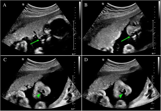
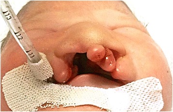
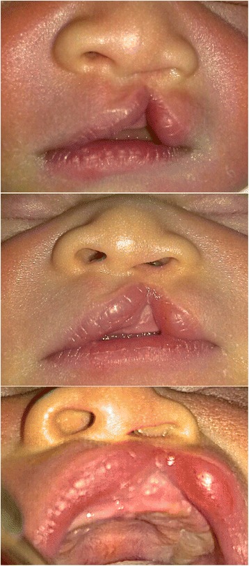
Ultrasonographic examination is widely used for screening of abnormal findings on prenatal screening. Cleft lip with or without cleft palate of the fetus can also be screened by using ultrasonography. Presence of abnormal findings of the fetal lip or palate can be detected by the imaging professionals. However, such findings may not be familiar to oral and maxillofacial surgeons. Oral and maxillofacial surgeons can use ultrasonographic imaging of fetal cleft lip with or without cleft palate to provide information regarding treatment protocols and outcomes to the parent. Therefore, surgeons should also be able to identify the abnormal details from the images, in order to setup proper treatment planning after the birth of the fetus. We report two cases of cleft lip with or without cleft palate that the official readings of prenatal ultrasonography were inconsistent with the actual facial structure identified after birth. Also, critical and practical points in fetal ultrasonographic diagnosis are to be discussed.
Keywords: Cleft lip; Cleft palate; Prenatal ultrasonography.
Figures

Similar articles
-
Ultrasound diagnosis of cleft lip and cleft palate before birth.Plast Reconstr Surg. 1981 Dec;68(6):854-9. doi: 10.1097/00006534-198112000-00002. Plast Reconstr Surg. 1981. PMID: 7301980
-
Fetal cleft lip and palate detection by three-dimensional ultrasonography.Ultrasound Obstet Gynecol. 2000 Sep;16(4):314-20. doi: 10.1046/j.1469-0705.2000.00181.x. Ultrasound Obstet Gynecol. 2000. PMID: 11169306
-
Fetal cleft lip with and without cleft palate: comparison between MR imaging and US for prenatal diagnosis.Eur J Radiol. 2011 Sep;79(3):437-42. doi: 10.1016/j.ejrad.2010.03.026. Epub 2010 Apr 24. Eur J Radiol. 2011. PMID: 20418035
-
[Prenatal diagnosis of cleft lip with or without cleft palate: retrospective study and review].J Gynecol Obstet Biol Reprod (Paris). 2013 Apr;42(2):151-8. doi: 10.1016/j.jgyn.2012.08.002. Epub 2012 Sep 25. J Gynecol Obstet Biol Reprod (Paris). 2013. PMID: 23017738 Review. French.
-
In utero surgery for cleft lip/palate: minimizing the "Ripple Effect" of scarring.J Craniofac Surg. 2003 Jul;14(4):504-11. doi: 10.1097/00001665-200307000-00021. J Craniofac Surg. 2003. PMID: 12867864 Review.
Cited by
-
Fistula incidence after primary repair and correlation with cleft width-to-palatum width ratio: A prospective cohort study.Ann Med Surg (Lond). 2021 Feb 23;63:102183. doi: 10.1016/j.amsu.2021.102183. eCollection 2021 Mar. Ann Med Surg (Lond). 2021. PMID: 33717475 Free PMC article.
-
The Importance of Multidisciplinary Management during Prenatal Care for Cleft Lip and Palate.Arch Plast Surg. 2016 Mar;43(2):153-9. doi: 10.5999/aps.2016.43.2.153. Epub 2016 Mar 18. Arch Plast Surg. 2016. PMID: 27019808 Free PMC article.
-
Prevalence of non-syndromic orofacial clefts: based on 15,094,978 Chinese perinatal infants.Oncotarget. 2018 Jan 13;9(17):13981-13990. doi: 10.18632/oncotarget.24238. eCollection 2018 Mar 2. Oncotarget. 2018. PMID: 29568410 Free PMC article.
References
-
- Jones MC (2002) Prenatal diagnosis of cleft lip and palate: detection rates, accuracy of ultrasonography, associated anomalies, and strategies for counseling. Cleft Palate Craniofac J 39:169–173, http://dx.doi.org/10.1597/1545-1569(2002)039<0169:PDOCLA>2.0.CO;2 - PubMed
-
- Maarse W, Berge SJ, Pistorius L, van Barneveld T, Kon M, Breugem C, Mink van der Molen AB (2010) Diagnostic accuracy of transabdominal ultrasound in detecting prenatal cleft lip and palate: a systematic review. Ultrasound Obstet Gynecol 35:495–502. doi:10.1002/uog.7472 - PubMed
Publication types
LinkOut - more resources
Full Text Sources
Other Literature Sources