. 2015 Oct;21(10):1209-15.
doi: 10.1038/nm.3931. Epub 2015 Aug 31.

CD47 blockade triggers T cell-mediated destruction of immunogenic tumors

Affiliations

CD47 blockade triggers T cell-mediated destruction of immunogenic tumors

Xiaojuan Liu et al. Nat Med. 2015 Oct.

Abstract

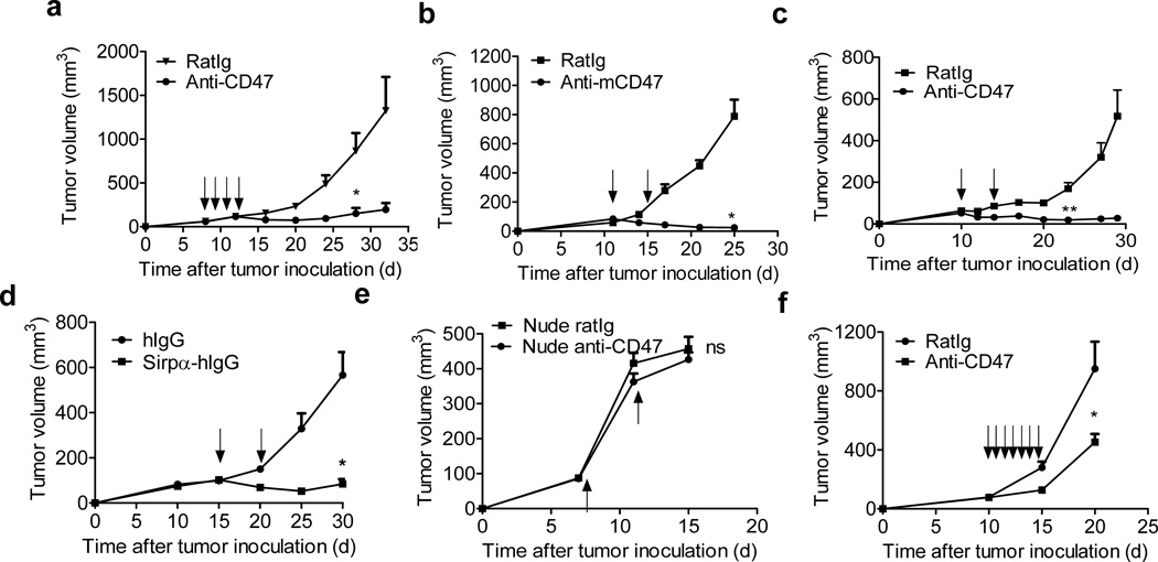
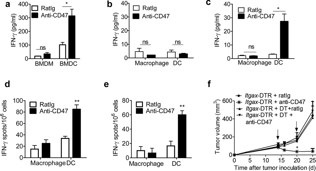
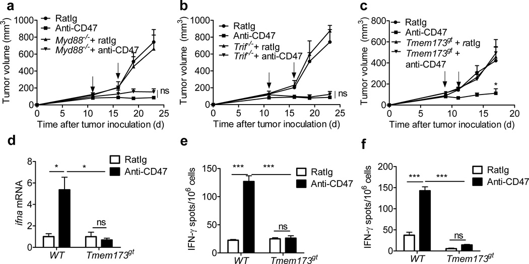
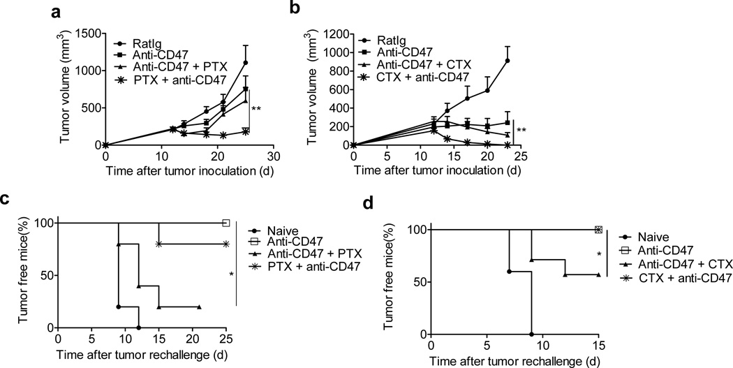
Macrophage phagocytosis of tumor cells mediated by CD47-specific blocking antibodies has been proposed to be the major effector mechanism in xenograft models. Here, using syngeneic immunocompetent mouse tumor models, we reveal that the therapeutic effects of CD47 blockade depend on dendritic cell but not macrophage cross-priming of T cell responses. The therapeutic effects of anti-CD47 antibody therapy were abrogated in T cell-deficient mice. In addition, the antitumor effects of CD47 blockade required expression of the cytosolic DNA sensor STING, but neither MyD88 nor TRIF, in CD11c+ cells, suggesting that cytosolic sensing of DNA from tumor cells is enhanced by anti-CD47 treatment, further bridging the innate and adaptive responses. Notably, the timing of administration of standard chemotherapy markedly impacted the induction of antitumor T cell responses by CD47 blockade. Together, our findings indicate that CD47 blockade drives T cell-mediated elimination of immunogenic tumors.


Figures

Figure 1
Figure 1. Anti-tumor effects of anti-CD47 depend on T cells
(a) Balb/c mice (n=5/group) transplanted s.c. with 5×10⁶ A20 cells were treated intraperitoneally with 400µg of anti-mCD47or isotype control rat Ig on days 7, 9, 11 and 13. (b) BALB/c mice (n=5/group) were injected s.c. with 5×10⁶ A20 cells and treated intratumorally with 50µg of anti-CD47 or isotype control rat Ig on days 11 and 16. (c) C57BL/6 mice (n=5/group) were injected s.c. with 106 MC38 cells and treated intratumorally with 50µg of anti-CD47 or isotype control rat Ig on days 10 and 14. One of three representative experiments is shown. (d) A20 tumor-bearing Balb/c mice (n=5/group) were injected s.c. with 5×10⁶ A20 cells and treated twice with 50µg of Sirpα-hIg or human intratumorally on days 15 and 20 (e) Balb/c nude mice (n=4/group) were injected s.c. with 2×10⁶ A20 cells and intratumorally injected with 50µg of anti-CD47 or isotype control rat Ig on days 7 and 11. (f) 2×10⁶ A20 were transplanted subcutaneously on Balb/c nude mice. When tumors were established (> 50 mm3), treatment began with daily intratumoral injections of 50 µg anti-CD47or 50 µg rat Ig for one week since day 10. Tumor growth is reported as the mean tumor size ±s.e.m. over time. One representative experiment out of three (b–c) or two (a, d-f) yielding similar results is depicted. ns (nonsignificant), *p < 0.05, **P < 0.01 (unpaired Student's t test) compared to tumors treated with RatIg.
Figure 2
Figure 2. Therapeutic effect of anti-CD47 requires CD8+ T cells
(a) BALB/c (n=8/group) mice were injected s.c. with 5×10⁶ A20 and treated intratumorally with 50µg of anti-CD47 or rat Ig on days 11 and 15. 200 µg of CD8 or CD4-depleting antibody was administered twice a week, starting on day 11. (b) Tumor-free, antibody-treated Balb/c mice (n=7/group) were rechallenged s.c. with 2.5×10⁷ A20 cells on opposite site from primary tumor one month after complete rejection. (c) MC38-OTI tumor bearing (n=6/group) B6 mice were i.t. treated twice with 50µg of either anti-CD47 or rat Ig on days 11 and 14. After 5 days, lymphocytes from draining LNs were isolated and stimulated with 10µg/ml OTI peptide. IFN-γ producing cells were enumerated by ELISPOT assay. (d) Balb/c mice (n=3) were injected s.c. with 5×10⁶ A20 and i.t. treated with 50 µg of either anti-CD47 or rat Ig antibody on days 11 and 16. 7 days after the final treatment, 2×10⁵ DLN cells from mice treated with anti-CD47 or rat Ig were stimulated with A20 tumor cells irradiated with 60Gy. The ratio of DLN cells to irradiated tumor was 5:1. IFN-γ -producing cells were enumerated by ELISPOT assay. (e) A20 tumor-bearing mice (n=5/group) were treated twice with 50µg of anti-mCD47 or rat Ig intratumorally on days 8 and 13. 300µg anti-IFNγ or rat Ig isotype control were injected intraperitoneally every four days. Data are reported as the means ±s.e.m. over time. *p < 0.05 **p < 0.01 ***p < 0.001 (unpaired Student's t test). One of three independent experiments is shown.
Fig3
Fig3. Anti-CD47 triggers the cross-priming ability of DCs
(a) BMDCs or BMM were cultured with MC38-OTIp in the presence of fresh GM-CSF and anti-CD47 overnight. Subsequently purified CD11c+ cells or F4/80+ cells were co-cultured with isolated CD8+ T cells from naive OTI mice for three days and analyzed by IFN-γ CBA. (b)–(c) MC38-OT1p bearing mice (n=5/group) were treated twice with 50µg of either anti-CD47 or rat Ig on days 11 and 14 intratumorally. Five days after the initial treatment, DLN (b) and Tumor (c) infiltrating DCs and macrophages were isolated and co-cultured with isolated CD8+ T cells from naive OTI mice for three days. IFN-γ production was detected by CBA. (d) A20 or (e) MC38 tumor bearing B6 mice (n=5/group) were treated with 50 µg of either anti-CD47 or rat Ig isotype control on day 10. Four days after the antibody treatment, 3×10⁴ Tumor infiltrating DCs and macrophages were isolated and co-cultured with isolated 3×10⁵ CD8+ T cells from A20 (d) or MC38 (e) vaccined-mice. 48 hours later, IFN-γ-producing cells were enumerated by ELISPOT assay. (f) 5 weeks after the indicated CD11c–DTR bone marrow chimera reconstitution, B6 mice (n=5/group) were injected subcutaneously with 106 MC38 cells and treated with 50µg of anti-CD47 or rat Ig on days 14 and 20. Diphtheria toxin or PBS was administrated on the same day as treatment. Data are reported as the means ±s.e.m.. *p < 0.05 **p < 0.01 ***p < 0.001 (unpaired Student's t test). One representative experiment out of three independent experiments is depicted.
Figure4
Figure4. Type I IFNs are induced during anti-CD47 mediated tumor Inhibition and required
(a) Four days after anti-CD47 or Rat Ig treatment (n=5/group), the single cell suspensions from tumors were sorted into CD45+CD11c CD11b+Ly6chi(Monocytes), CD45+CD11c−/+ CD11b+Ly6c F4/80+(Macrophage) and CD45+CD11c+CD11b−/+Ly6C F4/80 (DC)populations. mRNA level of ifna and ifnb in different cell subsets were quantified by real-time PCR assay. Representative data are reported as mean copy numbers ±s.e.m. after intrasample normalization to the levels of reference gene hprt in three independent experiments. *p < 0.05, **p<0.01, ***p < 0.001 (unpaired Student's t test). (b) C57BL/6 mice (n = 6) were injected s.c. with 106 MC38 cells and treated intratumorally with 50µg of anti-CD47 or isotype control rat Ig on days 11 and 14. 50µg anti-IFNAR1 antibody was administered intratumorally on days 0 and 2 after anti-CD47 treatment. (c)Ifnar1flox/flox and CD11c–CreIfnar1flox/flox mice (n = 6) were injected s.c. with 106 MC38 cells and treated with 50µg of anti-CD47 or rat Ig on days 10 and 15. (d) BMDCs from WT mice or ifnar1−/− mice were cultured with MC38-OTIp in the presence of anti-CD47 or rat Ig for 16h. Subsequently purified CD11c+ cells were cocultured with isolated CD8+ T cells from naive OT-I mice for 3 days. IFN-γ production in supernatant was determined by CBA. Data are reported as means ±s.e.m. Tumor growth is reported as the mean tumor size ±s.e.m. over time. **p < 0.01; One representative experiment out of three independent experiments is depicted.
Figure5
Figure5. STING signaling is required for anti-CD47 mediated tumor inhibition
MC38 tumors established (> 50mm3) in mice were treated i.t. with anti-CD47 or rat Ig. (a) Tumor growth in WT and Myd88−/− mice (n=5/group) (b) Tumor growth in WT and Trif−/− mice (n=5/group) (c) Tumor growth in WT and Tmem173gt mice (n=6/group) (d) WT mice or Tmem173gt mice (n=5/group) were injected s.c. with MC38 cells and i.t. treated with 50µg of anti-CD47 on days 12 and 15. Five days after the initial treatment, DCs were sorted from tumors. mRNA level of ifna were quantified by real-time PCR assay. Representative data are reported as mean copy numbers ±s.e.m. after intrasample normalization to the levels of hprt in three independent experiments. *p < 0.05 (unpaired Student's t test). (e) BMDCs from WT mice or Tmem173gtmice were cultured with MC38-OTI in the presence of anti-CD47 or rat Ig for 16h. Subsequently purified CD11c+ cells were cocultured with purifed OT-I cells for 2 days. IFN-γ production was determined by ELISPOT assay. (f) MC38 tumor bearing B6 mice (n=5/group) were sacrificed 7 days after the final treatment. 2.5×10⁵ CD8+ T cells, isolated from DLNs, were stimulated with MC38 tumor cells. The ratio of CD8+ T cells to MC38 was 50:1. IFN-γ-producing cells were enumerated by ELISPOT assay. Result was expressed as number of spots per 106 CD8+ T cells. Data are reported as means ±s.e.m. *p < 0.05; **p < 0.01; ***p < 0.001(unpaired Student's t test); One of three experiments is shown.
Figure 6
Figure 6. Anti-CD47-mediated immune protection is impaired by some post treatment Chemotherapeutics
Balb/c mice (n = 6/group) were injected s.c. with 3×10⁶ A20 cells and treated with 50µg of anti-CD47 on days 12 and 17. Select chemotherapeutic agents were injected i.p. at different time points. (a) 40 mg/kg of PTX was injected i.p. with single dose on day 11 (one day before anti-CD47) or three doses on days 15, 18 and 21 (since 3 days post anti-CD47) (n = 7–9 pooled from two experiments). Tumor growth is reported as the mean tumor size ±s.e.m over time. **p < 0.01 (unpaired Student's t test). (b) 60 mg/kg of CTX (n=5/group) was injected i.p. with single dose on day 11 (one day before anti-CD47) or three doses on days 15, 18 and 21(since 3 days post anti-CD47). Tumor growth is reported as the mean tumor size ±s.e.m over time. **p < 0.01 (Two-way ANOVA). One representative experiment out of two independent experiments is depicted. (c–d) Treated mice (n=7/group) were removed tumors by surgery and rechallenged with 1.5×10⁷ A20 cells one week after surgery. Percentage of tumor-free mice is shown. *p < 0.05 (Mantel-Cox). One representative experiment out of two independent experiments is depicted.

Comment in

References

    1. Gardai SJ, et al. Cell-surface calreticulin initiates clearance of viable or apoptotic cells through trans-activation of LRP on the phagocyte. Cell. 2005;123:321–334. - PubMed
    1. Chao MP, et al. Calreticulin is the dominant pro-phagocytic signal on multiple human cancers and is counterbalanced by CD47. Sci Transl Med. 2010;2:63ra94. - PMC - PubMed
    1. Chao MP, Majeti R, Weissman IL. Programmed cell removal: a new obstacle in the road to developing cancer. Nat Rev Cancer. 2012;12:58–67. - PubMed
    1. Oldenborg PA, et al. Role of CD47 as a marker of self on red blood cells. Science. 2000;288:2051–2054. - PubMed
    1. Blazar BR, et al. CD47 (integrin-associated protein) engagement of dendritic cell and macrophage counterreceptors is required to prevent the clearance of donor lymphohematopoietic cells. J Exp Med. 2001;194:541–549. - PMC - PubMed

Publication types