. 2015 Aug 31;10(9):e0134946.
doi: 10.1371/journal.pone.0134946. eCollection 2015.

Differential Expression of Specific Dermatan Sulfate Domains in Renal Pathology

Affiliations

Differential Expression of Specific Dermatan Sulfate Domains in Renal Pathology

Joost F M Lensen et al. PLoS One. .

Abstract

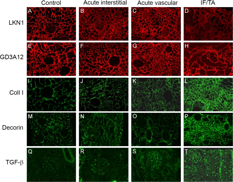
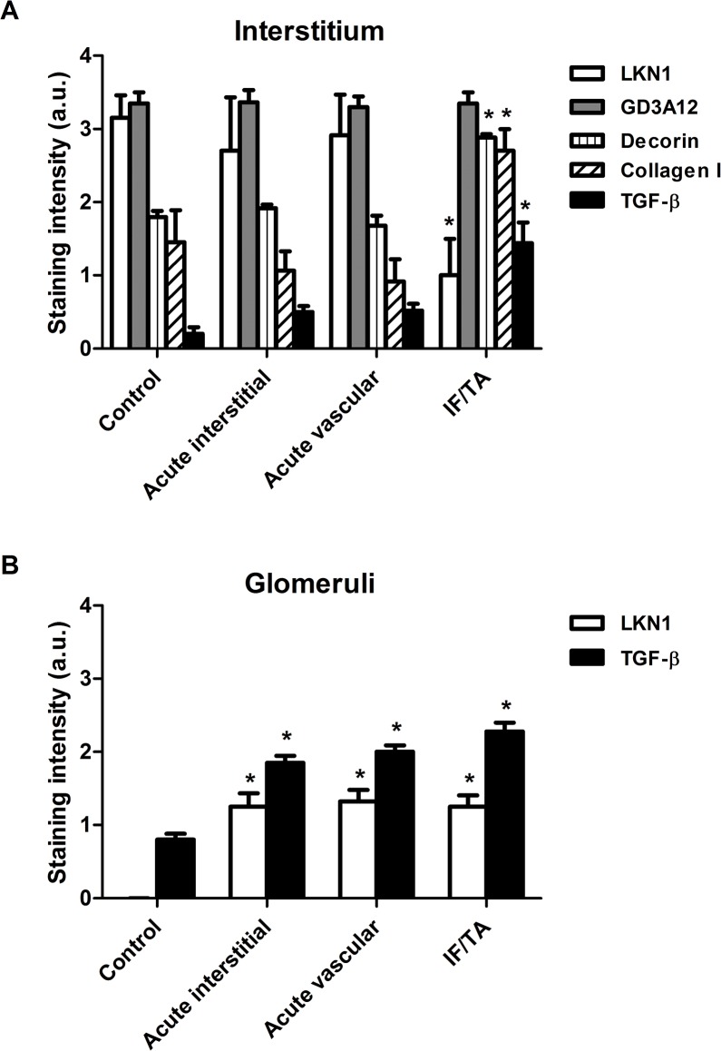
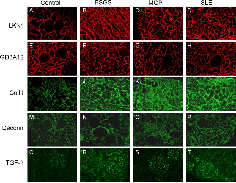
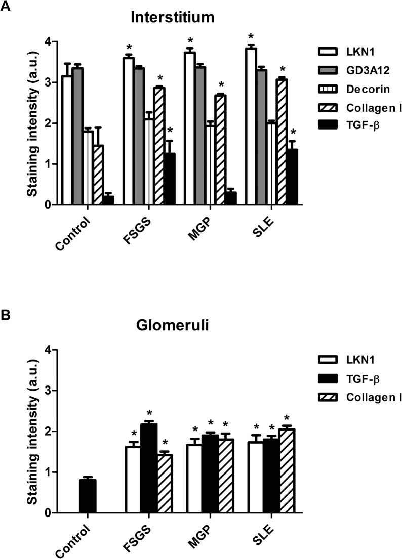
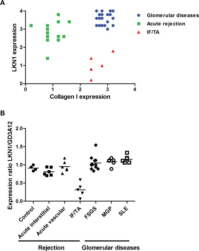
Dermatan sulfate (DS), also known as chondroitin sulfate (CS)-B, is a member of the linear polysaccharides called glycosaminoglycans (GAGs). The expression of CS/DS and DS proteoglycans is increased in several fibrotic renal diseases, including interstitial fibrosis, diabetic nephropathy, mesangial sclerosis and nephrosclerosis. Little, however, is known about structural alterations in DS in renal diseases. The aim of this study was to evaluate the renal expression of two different DS domains in renal transplant rejection and glomerular pathologies. DS expression was evaluated in normal renal tissue and in kidney biopsies obtained from patients with acute interstitial or vascular renal allograft rejection, patients with interstitial fibrosis and tubular atrophy (IF/TA), and from patients with focal segmental glomerulosclerosis (FSGS), membranous glomerulopathy (MGP) or systemic lupus erythematosus (SLE), using our unique specific anti-DS antibodies LKN1 and GD3A12. Expression of the 4/2,4-di-O-sulfated DS domain recognized by antibody LKN1 was decreased in the interstitium of transplant kidneys with IF/TA, which was accompanied by an increased expression of type I collagen, decorin and transforming growth factor beta (TGF-β), while its expression was increased in the interstitium in FSGS, MGP and SLE. Importantly, all patients showed glomerular LKN1 staining in contrast to the controls. Expression of the IdoA-Gal-NAc4SDS domain recognized by GD3A12 was similar in controls and patients. Our data suggest a role for the DS domain recognized by antibody LKN1 in renal diseases with early fibrosis. Further research is required to delineate the exact role of different DS domains in renal fibrosis.


Conflict of interest statement

Competing Interests: The authors have declared that no competing interests exist.

Figures

Fig 1
Fig 1. Expression of the 4/2,4-di-O-sulfated and IdoA-Gal-NAc4S DS domains defined by the antibodies LKN1 (A-D) and GD3A12 (E-H), type I collagen (I-L), decorin (M-P) and TGF-β (Q-T) in renal allograft rejection and controls.
Representative photographs showing the expression of the 4/2,4-di-O-sulfated DS domain defined by LKN1 in the control human kidneys (A). Tubular interstitial expression of this 4/2,4-di-O-sulfated DS domain defined by LKN1 was increased in acute interstitial (B) and acute vascular (C) renal allograft rejections compared to interstitial fibrosis and tubular atrophy (IF/TA) (D). Expression of the IdoA-Gal-NAc4S DS domain recognized by GD3A12 was similar in the control human kidney and the three types of renal allograft rejection (E-H), while expression of type I collagen (coll I) and decorin was increased in IF/TA (L, P) compared to the control human kidney (I, M), and acute interstitial (J, N) and acute vascular renal allograft rejections (K, O). Glomerular expression of transforming growth factor beta (TGF-β) was increased in the three types of renal allograft rejection (R-T) compared to the control human kidney (Q). Magnification A-P 100x, magnification Q-T 200x.
Fig 2
Fig 2. Semi-quantitative analysis of the expression of type I collagen, decorin, TGF-β and the 4/2,4-di-O-sulfated and IdoA-Gal-NAc4S DS domains defined by LKN1 and GD3A12 in the interstitium (A) and glomeruli (B) in renal allograft rejection.
Staining intensities of type I collagen, decorin, transforming growth factor beta (TGF-β) and the DS domains defined by LKN1 and GD3A12 were scored using a scale of 0–4 and revealed a significantly increased tubular interstitial expression of type I collagen, decorin and TGF-β and a decreased expression of the 4/2,4-di-O-sulfated DS domain recognized by LKN1 in interstitial fibrosis and tubular atrophy (IF/TA). Expression of the IdoA-Gal-NAc4S DS domain recognized by GD3A12 was similar in controls and the three types of renal allograft rejection (A). Glomerular expression of the 4/2,4-di-O-sulfated DS domain defined recognized by LKN1 and TGF-β was increased in the acute interstitial, acute vascular and chronic renal allograft rejections compared to controls (B). *P<0.05 vs control.
Fig 3
Fig 3. Expression of the 4/2,4-di-O-sulfated and IdoA-Gal-NAc4S DS domains defined by LKN1 (A-D) and GD3A12 (E-H), type I collagen (I-L), decorin (M-P) and TGF-β (Q-T) in renal sections obtained from patients with glomerular diseases.
Representative photographs showing the immunofluorescence stainings of renal sections obtained from controls (A, E, I, M, Q), patients with focal segmental glomerulosclerosis (FSGS) (B, F, J, N, R), membranous glomerulopathy (MGP) (C, G, K, O, S) and systemic lupus erythematosus (SLE) (D, H, L, P, T) with the anti-DS antibodies LKN1 (A-D) and GD3A12 (E-H), anti-collagen type I (Coll I) (I-L), decorin (M-P) and transforming growth factor beta (TGF-β) (Q-T). Tubular interstitial and glomerular expression of the 4/2,4-di-O-sulfated DS domain defined by LKN1 and of type I collagen was increased in FSGS (B, J), MGP (C, K) and SLE (D, L) compared to control human kidney (A, I). Expression of the IdoA-Gal-NAc4S DS domain, recognized by GD3A12, and decorin was similar in the interstitium and glomeruli of patients (F-H, N-P) and controls (E, M). Glomerular expression of TGF-β was increased in FSGS (R), MGP (S) and SLE (T) compared to control (Q). Magnification A-P 100x, magnification Q-T 200x.
Fig 4
Fig 4. Semi-quantitative analysis of the expression the 4/2,4-di-O-sulfated and IdoA-Gal-NAc4S DS domains defined by LKN1 and GD3A12, type I collagen, decorin and TGF-β in the interstitium (A) and glomeruli (B) of patients with FSGS, MGP and SLE.
Staining intensities of the 4/2,4-di-O-sulfated and IdoA-Gal-NAc4S DS domains defined by LKN1 and GD3A12 respectively and of type I collagen, decorin and transforming growth factor beta (TGF-β) were scored using a scale of 0–4 and revealed a significantly increased expression of the 4/2,4-di-O-sulfated DS domain and type I collagen in the interstitium (A) and glomeruli (B) of patients with glomerular diseases. Glomerular expression of TGF-β also was increased in patients with glomerular diseases (B), while expression of the IdoA-Gal-NAc4S DS domain and decorin was similar in the interstitium (A) of patients and controls. *P<0.05 vs control.
Fig 5
Fig 5. Correlation between the expression of the 4/2,4-di-O-sulfated DS domain recognized by antibody LKN1 and type I collagen (A) and the expression ratio of LKN1 and GD3A12 (B) in glomerular diseases, acute rejection and IF/TA.
In acute (vascular and interstitial) rejection, the interstitial expression of the 4/2,4-di-O-sulfated DS domain recognized by antibody LKN1 is increased, while expression of collagen type I is decreased (A). Expression of collagen type I is increased in glomerular diseases and IF/TA, which is accompanied by an increased expression of the 4/2,4-di-O-sulfated DS domain in glomerular diseases and a decreased expression of the 4/2,4-di-O-sulfated DS domain in IF/TA. The expression ratio of the 4/2,4-di-O-sulfated DS domain recognized by antibody LKN1 to the IdoA-Gal-NAc4S DS domain recognized by antibody GD3A12 is decreased in the IF/TA patients in contrast to the patients with acute rejection or glomerular diseases (B).

Similar articles

Cited by

References

    1. Malavaki C, Mizumoto S, Karamanos N, Sugahara K. Recent advances in the structural study of functional chondroitin sulfate and dermatan sulfate in health and disease. Connective tissue research. 2008;49(3):133–9. 10.1080/03008200802148546 - DOI - PubMed
    1. Trowbridge JM, Gallo RL. Dermatan sulfate: new functions from an old glycosaminoglycan. Glycobiology. 2002;12(9):117R–25R. - PubMed
    1. Sugahara K, Mikami T, Uyama T, Mizuguchi S, Nomura K, Kitagawa H. Recent advances in the structural biology of chondroitin sulfate and dermatan sulfate. Current opinion in structural biology. 2003;13(5):612–20. - PubMed
    1. Corsi A, Xu T, Chen XD, Boyde A, Liang J, Mankani M, et al. Phenotypic effects of biglycan deficiency are linked to collagen fibril abnormalities, are synergized by decorin deficiency, and mimic Ehlers-Danlos-like changes in bone and other connective tissues. Journal of bone and mineral research: the official journal of the American Society for Bone and Mineral Research. 2002;17(7):1180–9. - PubMed
    1. Danielson KG, Baribault H, Holmes DF, Graham H, Kadler KE, Iozzo RV. Targeted disruption of decorin leads to abnormal collagen fibril morphology and skin fragility. The Journal of cell biology. 1997;136(3):729–43. - PMC - PubMed

Publication types