. 2015 Sep;71(Pt 9):1125-30.
doi: 10.1107/S2053230X15012698. Epub 2015 Aug 25.

Structure of RizA, an L-amino-acid ligase from Bacillus subtilis

Affiliations

Structure of RizA, an L-amino-acid ligase from Bacillus subtilis

Wataru Kagawa et al. Acta Crystallogr F Struct Biol Commun. 2015 Sep.

Abstract

RizA is an L-amino-acid ligase from Bacillus subtilis that participates in the biosynthesis of rhizocticin, an oligopeptide antibiotic. The substrate-free form of RizA has been crystallized and the structure was solved at 2.8 Å resolution. The amino-acid-binding site appears to be capable of accommodating multiple amino acids, consistent with previous biochemical studies.

Keywords: ATP-grasp fold; dipeptide synthesis; l-amino-acid ligase; rhizocticin.


Figures

Figure 1
Figure 1
Overall structure of RizA. The RizA structure determined at 2.8 Å resolution is depicted as a ribbon representation. RizA has an ATP-grasp fold consisting of the A-, B- and C-domains (coloured blue, purple and yellow, respectively).
Figure 2
Figure 2
Comparison of the domain orientations in the RizA structure with those of other LALs. (ac) Structural superpositions of RizA (a) with BL00235 (b) and BacD (c). RizA is coloured according to the A-, B- and C-domains (blue, purple and yellow, respectively). BL00235 and BacD are coloured light cyan and light green, respectively. (df) Surface representations of RizA (d), BL00235 (e) and BacD (f). For the three proteins, the A-, B- and C-­domains are coloured blue, purple and yellow, respectively. ADP (coloured red), divalent ions (Mg2+ or Ca2+, coloured green) and the amino-acid substrate (BacD only, coloured dark blue) bound to BL00235 (g) and BacD (h) are viewed from the same angle as in (e) and (f), respectively.
Figure 3
Figure 3
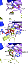
The putative ATP-binding site of RizA. The conserved amino-acid residues involved in ADP binding, revealed from the crystal structures of BacD and BL00235, are shown in stick representations. All three domains appear to contribute to ATP binding. (a) Lys111 (coloured blue) is located in the A-domain, Lys151 and Glu193 (coloured purple) are located in the B-domain, and Glu259 and Glu272 (coloured yellow) are located in the C-domain. The surrounding protein regions are shown in ribbon representations and are coloured according to the three domains. The green spheres in (b) and (c) depict Mg2+ and Ca2+ ions, respectively. Dashed lines (coloured light blue) indicate hydrogen-bonding or ionic interactions.
Figure 4
Figure 4
The putative dipeptide-binding site of RizA. The predicted dipeptide-binding site of RizA is depicted as surface (a) and ribbon (b) representations. The phosphorylated phosphinate l-alanyl-l-phenylalanine and ADP from the superpositioned BacD structure are shown. The A-domain (white dashed circle) is the predicted binding site for the arginine side chain. Several hydrophobic residues are located in this region that may interact with the alkyl chain of the arginine residue. In addition, the disordered amino-acid residues 222–230 (VPLLKGMAK) and 368–375 (DEKQSSSF) are near this region and contain several hydrophobic amino-acid residues that may interact with arginine. The carboxyl group of the Asp376 residue is coloured red. The B-domain (white dashed circle) is the predicted binding site for the C-terminal amino-acid residue of the dipeptide.

Similar articles

Cited by

References

    1. Adams, P. D. et al. (2010). Acta Cryst. D66, 213–221. - PubMed
    1. Arai, T., Arimura, Y., Ishikura, S. & Kino, K. (2013). Appl. Environ. Microbiol. 79, 5023–5029. - PMC - PubMed
    1. Arai, T. & Kino, K. (2008). Biosci. Biotechnol. Biochem. 72, 3048–3050. - PubMed
    1. Bender, C. L., Alarcón-Chaidez, F. & Gross, D. C. (1999). Microbiol. Mol. Biol. Rev. 63, 266–292. - PMC - PubMed
    1. Emsley, P., Lohkamp, B., Scott, W. G. & Cowtan, K. (2010). Acta Cryst. D66, 486–501. - PMC - PubMed

Publication types

Associated data