. 2015 Sep;71(Pt 9):1189-93.
doi: 10.1107/S2053230X15013990. Epub 2015 Aug 25.

Expression, purification, crystallization and X-ray diffraction studies of the molecular chaperone prefoldin from Homo sapiens

Affiliations

Expression, purification, crystallization and X-ray diffraction studies of the molecular chaperone prefoldin from Homo sapiens

Yoshiki Aikawa et al. Acta Crystallogr F Struct Biol Commun. 2015 Sep.

Abstract

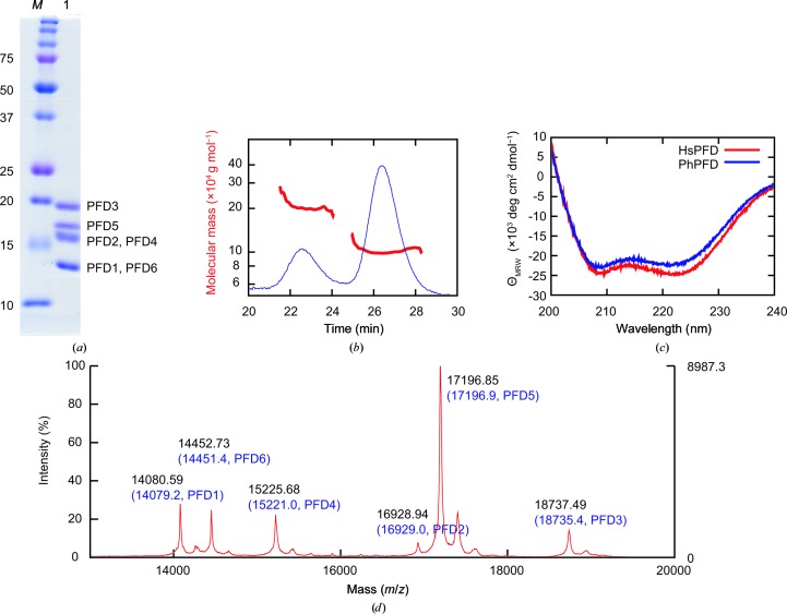
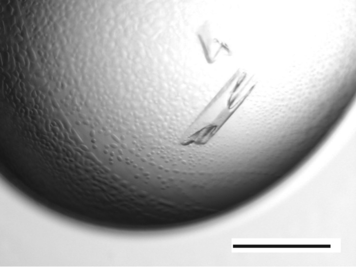
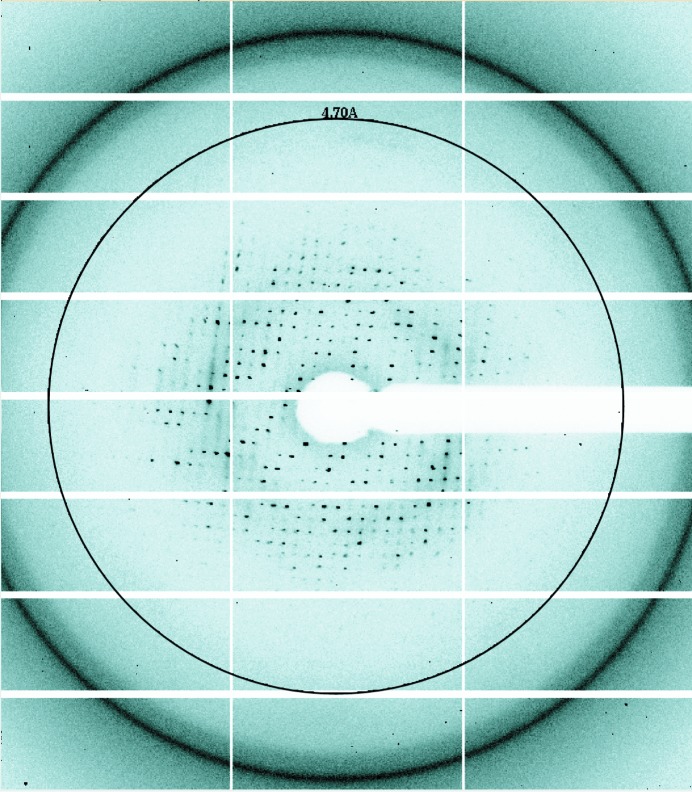
Proper protein folding is an essential process for all organisms. Prefoldin (PFD) is a molecular chaperone that assists protein folding by delivering non-native proteins to group II chaperonin. A heterohexamer of eukaryotic PFD has been shown to specifically recognize and deliver non-native actin and tubulin to chaperonin-containing TCP-1 (CCT), but the mechanism of specific recognition is still unclear. To determine its crystal structure, recombinant human PFD was reconstituted, purified and crystallized. X-ray diffraction data were collected to 4.7 Å resolution. The crystals belonged to space group P21212, with unit-cell parameters a = 123.2, b = 152.4, c = 105.9 Å.

Keywords: molecular chaperone; prefoldin; protein folding.


Figures

Figure 1
Figure 1
Biochemical characterization of human PFD. (a) SDS–PAGE analysis of purified human PFD. Lane M, molecular-weight marker (labelled in kDa); lane 1, peak fraction from Superdex 200 column. PFD subunits are assigned to the bands according to the molecular weights calculated from the sequences. (b) SEC-MALS of purified PFD. Red, molecular mass; blue, absorbance at 280 nm. (c) CD spectra. Red, spectrum of human PFD (HsPFD); blue, spectrum of P. horikoshii PFD (PhPFD). (d) MALDI-TOF MS. Peaks are annotated with the observed molecular mass (black) and the calculated mass from the sequence (blue, in parentheses).
Figure 2
Figure 2
Crystals of human PFD. The black bar represents 0.5 mm.
Figure 3
Figure 3
X-ray diffraction pattern from a crystal of human PFD. The circle represents 4.7 Å resolution.

References

    1. Hansen, W. J., Cowan, N. J. & Welch, W. J. (1999). J. Cell Biol. 145, 265–277. - PMC - PubMed
    1. Hartl, F. U. & Hayer-Hartl, M. (2002). Science, 295, 1852–1858. - PubMed
    1. Kida, H., Sugano, Y., Iizuka, R., Fujihashi, M., Yohda, M. & Miki, K. (2008). J. Mol. Biol. 383, 465–474. - PubMed
    1. Leroux, M. R., Fändrich, M., Klunker, D., Siegers, K., Lupas, A. N., Brown, J. R., Schiebel, E., Dobson, C. M. & Hartl, F. U. (1999). EMBO J. 18, 6730–6743. - PMC - PubMed
    1. Martín-Benito, J., Boskovic, J., Gómez-Puertas, P., Carrascosa, J. L., Simons, C. T., Lewis, S. A., Bartolini, F., Cowan, N. J. & Valpuesta, J. M. (2002). EMBO J. 21, 6377–6386. - PMC - PubMed

Publication types