

Long-term outcomes and prognostic factors of skull-base chondrosarcoma patients treated with pencil-beam scanning proton therapy at the Paul Scherrer Institute
- PMID: 26323608
- PMCID: PMC4724177
- DOI: 10.1093/neuonc/nov154
Long-term outcomes and prognostic factors of skull-base chondrosarcoma patients treated with pencil-beam scanning proton therapy at the Paul Scherrer Institute
Abstract
Background: Skull-base chondrosarcoma (ChSa) is a rare disease, and the prognostication of this disease entity is ill defined.
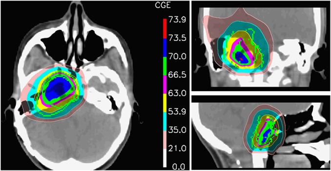
Methods: We assessed the long-term local control (LC) results, overall survival (OS), and prognostic factors of skull-base ChSa patients treated with pencil beam scanning proton therapy (PBS PT). Seventy-seven (male, 35; 46%) patients with histologically confirmed ChSa were treated at the Paul Scherrer Institute. Median age was 38.9 years (range, 10.2-70.0y). Median delivered dose was 70.0 GyRBE (range, 64.0-76.0 GyRBE). LC, OS, and toxicity-free survival (TFS) rates were calculated using the Kaplan Meier method.
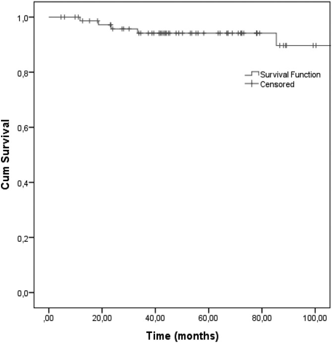
Results: After a mean follow-up of 69.2 months (range, 4.6-190.8 mo), 6 local (7.8%) failures were observed, 2 of which were late failures. Five (6.5%) patients died. The actuarial 8-year LC and OS were 89.7% and 93.5%, respectively. Tumor volume > 25 cm(3) (P = .02), brainstem/optic apparatus compression at the time of PT (P = .04) and age >30 years (P = .08) were associated with lower rates of LC. High-grade (≥3) radiation-induced toxicity was observed in 6 (7.8%) patients. The 8-year high-grade TFS was 90.8%. A higher rate of high-grade toxicity was observed for older patients (P = .073), those with larger tumor volume (P = .069), and those treated with 5 weekly fractions (P = .069).
Conclusions: This is the largest PT series reporting the outcome of patients with low-grade ChSa of the skull base treated with PBS only. Our data indicate that protons are both safe and effective. Tumor volume, brainstem/optic apparatus compression, and age were prognosticators of local failures.
Keywords: chondrosarcoma; pencil beam scanning; prognostic factors; proton therapy; skull-base tumors.
© The Author(s) 2015. Published by Oxford University Press on behalf of the Society for Neuro-Oncology. All rights reserved. For permissions, please e-mail: journals.permissions@oup.com.
Figures
References
-
- Oghalai JS, Buxbaum JL, Jackler RK, McDermott MW. Skull base chondrosarcoma originating from the petroclival junction. Otol Neurotol. 2005;26(5):1052–1060. - PubMed
-
- Rosenberg AE, Nielsen GP, Keel SB, et al. Chondrosarcoma of the base of the skull: a clinicopathologic study of 200 cases with emphasis on its distinction from chordoma. Am J Surg Pathol. 1999;23(11):1370–1378. - PubMed
MeSH terms
LinkOut - more resources
Full Text Sources
Other Literature Sources
Medical
