

Two-dimensional graphene as a matrix for MALDI imaging mass spectrometry
- PMID: 26323616
- PMCID: PMC4607658
- DOI: 10.1007/s13361-015-1243-6
Two-dimensional graphene as a matrix for MALDI imaging mass spectrometry
Abstract
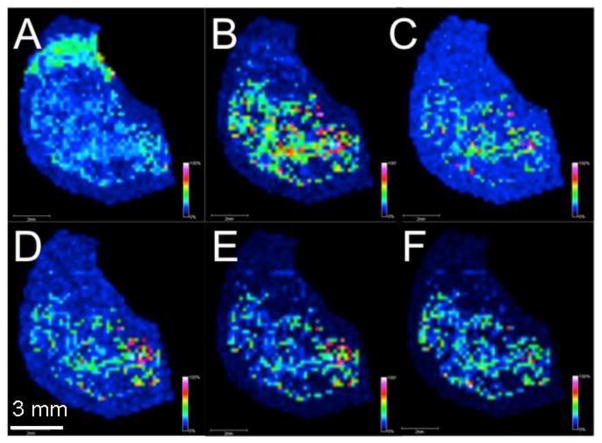
Here, a matrix using two-dimensional (2D) graphene is demonstrated for the first time in the context of MALDI IMS using a Fourier transform ion cyclotron resonance (FT-ICR) mass spectrometer. Although graphene flakes have been used previously in MALDI, it is described here how a single 2D layer of graphene is applied directly on top of rat brain sections and soybean leaves. Several classes of molecules are desorbed and ionized off of the surface of the tissues examined using 2D graphene, with minimal background interference from the matrix. Moreover, no solvents are employed in application of 2D graphene, eliminating the potential for analyte diffusion in liquid droplets during matrix application. Because 2D graphene is an elemental form of carbon, an additional advantage is its high compatibility with the long duration needed for many IMS experiments. Graphical Abstract ᅟ.
Keywords: Brain; Fourier transform ion cyclotron resonance; Graphene; Imaging mass spectrometry; MALDI; Soybean.
Figures


Similar articles
-
Mass spectrometry imaging of small molecules in biological tissues using graphene oxide as a matrix.Anal Chim Acta. 2017 Apr 15;962:52-59. doi: 10.1016/j.aca.2017.01.043. Epub 2017 Jan 31. Anal Chim Acta. 2017. PMID: 28231880
-
Simultaneous detection of phosphatidylcholines and glycerolipids using matrix-enhanced surface-assisted laser desorption/ionization-mass spectrometry with sputter-deposited platinum film.J Mass Spectrom. 2015 Nov;50(11):1264-9. doi: 10.1002/jms.3700. J Mass Spectrom. 2015. PMID: 26505771
-
Exploring three-dimensional matrix-assisted laser desorption/ionization imaging mass spectrometry data: three-dimensional spatial segmentation of mouse kidney.Anal Chem. 2012 Jul 17;84(14):6079-87. doi: 10.1021/ac300673y. Epub 2012 Jul 5. Anal Chem. 2012. PMID: 22720760
-
Matrix-assisted laser desorption/ionization imaging mass spectrometry: new technology for vascular pathology.J Vasc Res. 2014;51(2):144-8. doi: 10.1159/000362123. Epub 2014 May 8. J Vasc Res. 2014. PMID: 24820659 Review.
-
[Imaging Mass Spectrometry in Histopathologic Analysis].Rinsho Byori. 2015 Apr;63(4):472-80. Rinsho Byori. 2015. PMID: 26536781 Review. Japanese.
Cited by
-
Recent advances in mass spectrometry analysis of neuropeptides.Mass Spectrom Rev. 2023 Mar;42(2):706-750. doi: 10.1002/mas.21734. Epub 2021 Sep 24. Mass Spectrom Rev. 2023. PMID: 34558119 Free PMC article. Review.
-
Feasibility Assessment of a MALDI FTICR Imaging Approach for the 3D Reconstruction of a Mouse Lung.J Am Soc Mass Spectrom. 2017 Aug;28(8):1709-1715. doi: 10.1007/s13361-017-1658-3. Epub 2017 Apr 11. J Am Soc Mass Spectrom. 2017. PMID: 28401432
-
Large-Area Graphene Films as Target Surfaces for Highly Reproducible Matrix-Assisted Laser Desorption Ionization Suitable for Quantitative Mass Spectrometry.J Am Soc Mass Spectrom. 2018 Oct;29(10):2003-2011. doi: 10.1007/s13361-018-2024-9. Epub 2018 Jul 11. J Am Soc Mass Spectrom. 2018. PMID: 29998363
-
High-Throughput Metabolic Profiling of Soybean Leaves by Fourier Transform Ion Cyclotron Resonance Mass Spectrometry.Anal Chem. 2016 Jan 19;88(2):1188-94. doi: 10.1021/acs.analchem.5b03340. Epub 2015 Dec 23. Anal Chem. 2016. PMID: 26651857 Free PMC article.
-
A Rapid Screening Method for the Detection of Additives in Electronics and Plastic Consumer Products Using AP-MALDI-qTOF-MS.Toxics. 2023 Jan 23;11(2):108. doi: 10.3390/toxics11020108. Toxics. 2023. PMID: 36850984 Free PMC article.
References
-
- Zaima N, Matsuyama Y, Setou M. Principal component analysis of direct matrix-assisted laser desorption/ionization mass spectrometric data related to metabolites of fatty liver. Journal of oleo science. 2009;58:267–273. - PubMed
-
- Kruse R, Sweedler JV. Spatial profiling invertebrate ganglia using maldi ms. Journal of the American Society for Mass Spectrometry. 2003;14:752–759. - PubMed
-
- Aerni HR, Cornett DS, Caprioli RM. Automated acoustic matrix deposition for maldi sample preparation. Analytical chemistry. 2006;78:827–834. - PubMed
Publication types
MeSH terms
Substances
Grants and funding
LinkOut - more resources
Full Text Sources
Other Literature Sources
