Review
. 2015 Sep 26;370(1678):20140326.
doi: 10.1098/rstb.2014.0326.

Diversity and origins of anaerobic metabolism in mitochondria and related organelles

Affiliations
Review

Diversity and origins of anaerobic metabolism in mitochondria and related organelles

Courtney W Stairs et al. Philos Trans R Soc Lond B Biol Sci. .

Abstract

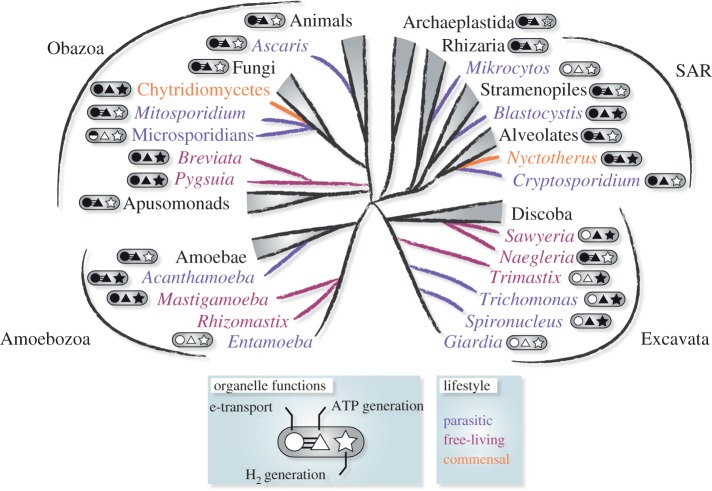
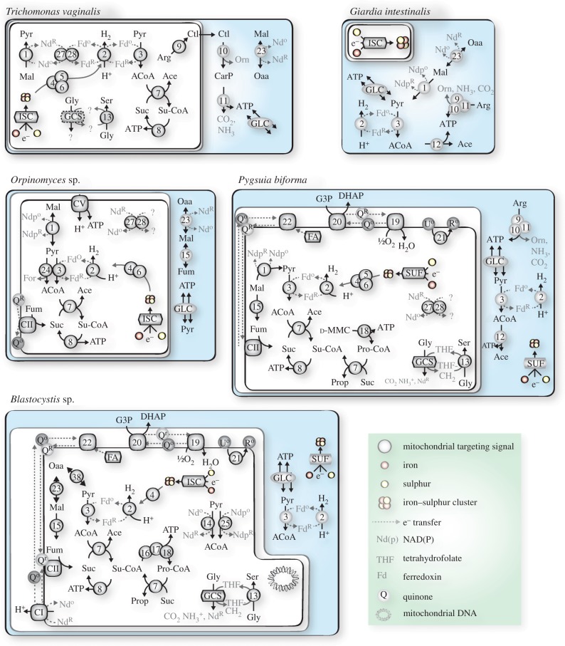
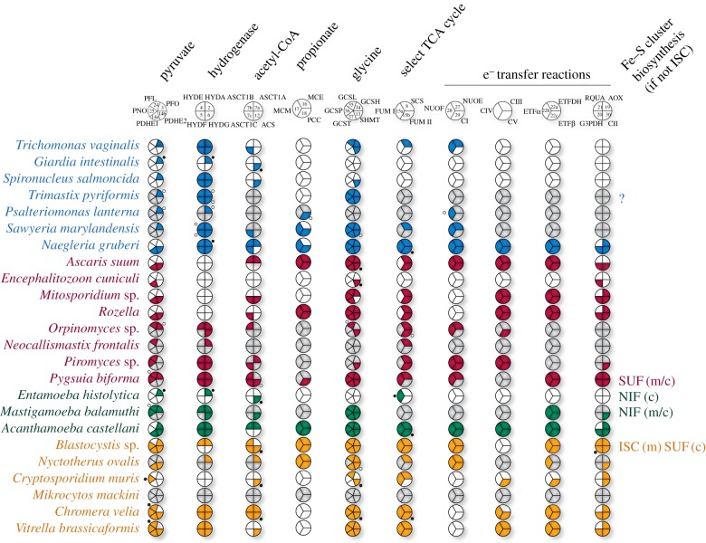
Across the diversity of life, organisms have evolved different strategies to thrive in hypoxic environments, and microbial eukaryotes (protists) are no exception. Protists that experience hypoxia often possess metabolically distinct mitochondria called mitochondrion-related organelles (MROs). While there are some common metabolic features shared between the MROs of distantly related protists, these organelles have evolved independently multiple times across the breadth of eukaryotic diversity. Until recently, much of our knowledge regarding the metabolic potential of different MROs was limited to studies in parasitic lineages. Over the past decade, deep-sequencing studies of free-living anaerobic protists have revealed novel configurations of metabolic pathways that have been co-opted for life in low oxygen environments. Here, we provide recent examples of anaerobic metabolism in the MROs of free-living protists and their parasitic relatives. Additionally, we outline evolutionary scenarios to explain the origins of these anaerobic pathways in eukaryotes.

Keywords: anaerobic metabolism; eukaryotic evolution; mitochondrion-related organelles.


Figures

Figure 1.
Figure 1.
Distribution of MROs across the major supergroups of eukaryotes. Organisms with parasitic (purple), commensal (orange) or free-living (red) lifestyles are indicated. Metabolic functions of each organism's MRO are indicated: shaded shapes represent the presence of electron transporting complexes (circle), ATP synthesis (triangle) and hydrogen production (star). Where at least one proton-pumping complex (CI, CIII, CIV) and ATP synthase (CV) was identified, the circle and triangle are joined by three lines. ‘*’ represents algal lineages where hydrogen production is located in the plastid.
Figure 2.
Figure 2.
Major metabolic pathways in the MROs of selected anaerobic protists. Oaa, oxaloacetate; Mal, malate; Pyr, pyruvate; Fum, fumarate; ACoA, acetyl-CoA; Ace, acetate; Suc, succinate; Su-CoA, succinyl-CoA; d-MMC, d-methylmalonyl-CoA; Prop, propionate; Pro-CoA, propionyl-CoA; Arg, arginine; Ctl, citrulline; Orn, ornithine; CarP, carbamoyl phosphate; Gly, glycine; Ser, serine. GLC, glycolysis; GCS, glycine cleavage system; ISC, iron–sulfur cluster system of ISC assembly; SUF, sulfur assimilation (SUF) system of ISC assembly; NIF, nitrogen fixation system of ISC assembly. THF, tetrahydrofolic acid. For the following, O: oxidized, R: reduced: Fd, ferredoxin; Nd, nicotinamide adenine dinucleotide (NAD+); Ndp, nicotinamide adenine dinucleotide phosphate (NADP+); Q, quinone; U, ubiquinone; R, rhodoquinone. 1, malic enzyme; 2, [FeFe]-hydrogenase (HYDA); 3, pyruvate : ferredoxin oxidoreductase (PFO); 4–6, hydrogenase maturases HydE, F G; 7, acetate : succinate CoA transferase (ASCT); 8, succinyl-CoA synthetase (SCS); 9, arginine deiminase (ADI); 10, ornithine transcarbamylase (OTC); 11, carbamate kinase (CK); 12, acetyl-CoA synthetase (ACS); 13, serine hydroxymethyltransferase (SHMT); 14, pyruvate dehydrogenase (PDH); 15, fumarase (FUM); 16, methylmalonyl-CoA epimerase (MCE); 17, methylmalonyl-CoA mutase (MCM); 18, propionyl-CoA carboxylase (PCC); 19, alternative oxidase (AOX); 20, glycerol-3-phosphate dehydrogenase (G3PDH); 21, rhodoquinone biosynthesis protein RQUA; 22, electron transport flavoprotein (ETF); 23, malate dehydrogenase (MDH); 24, pyruvate formate lyase (PFL); 25, pyruvate : NADP+ oxidoreductase (PNO); 27, NuoE/24 kDa subunit of ETC CI; 28, NuoF/51 kDa subunit of ETC CI; CI, complex I; CII, complex II (at least A and B subunits); CV, complex V.
Figure 3.
Figure 3.
Coulson plots showing the presence and absence of various proteins and/or protein subunits in anaerobic protists. Blue, Excavata; red, Obazoa; green, Amoebozoa; yellow, SAR clade (Stramenopiles, Alveolata and Rhizaria). Coloured sections indicate proteins found to be present in genome or transcriptome data; white sections, proteins absent from complete genome data; grey sections, proteins absent from transcriptome or incomplete genome data [19]. Black and white circles indicate proteins with a predicted cytosolic location based on Mitoprot predictions or incomplete coding sequences. Unless indicated otherwise, all lineages use the ISC system for the biosynthesis of Fe–S clusters. Enzyme abbreviations are as indicated in figures 2. Accession numbers for each protein can be found in the electronic supplementary material, S1.

Similar articles

Cited by

References

    1. Andersson SG, et al. 1998. The genome sequence of Rickettsia prowazekii and the origin of mitochondria. Nature 396, 133–140. (10.1038/24094) - DOI - PubMed
    1. Thrash JC, et al. 2011. Phylogenomic evidence for a common ancestor of mitochondria and the SAR11 clade. Sci. Rep. 1, 13 (10.1038/srep00013) - DOI - PMC - PubMed
    1. Rodríguez-Ezpeleta N, Embley TM. 2012. The SAR11 group of alpha-proteobacteria is not related to the origin of mitochondria. PLoS ONE 7, e30520 (10.1371/journal.pone.0030520) - DOI - PMC - PubMed
    1. Poole AM, Gribaldo S. 2014. Eukaryotic origins: how and when was the mitochondrion acquired? Cold Spring Harb. Perspect. Biol. 6, a015990 (10.1101/cshperspect.a015990) - DOI - PMC - PubMed
    1. Calvo SE, Mootha VK. 2010. The mitochondrial proteome and human disease. Annu. Rev. Genomics Hum. Genet. 11, 25–44. (10.1146/annurev-genom-082509-141720) - DOI - PMC - PubMed

Publication types