Clival osteomyelitis and hypoglossal nerve palsy--rare complications of Lemierre's syndrome
- PMID: 26323975
- PMCID: PMC4693136
- DOI: 10.1136/bcr-2015-209777
Clival osteomyelitis and hypoglossal nerve palsy--rare complications of Lemierre's syndrome
Abstract
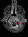
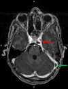
An increasingly reported entity, Lemierre's syndrome classically presents with a recent oropharyngeal infection, internal jugular vein thrombosis and the presence of anaerobic organisms such as Fusobacterium necrophorum. The authors report a normally fit and well 17-year-old boy who presented with severe sepsis following a 5-day history of a sore throat, myalgia and neck stiffness requiring intensive care admission. Blood cultures grew F. necrophorum and radiological investigations demonstrated left internal jugular vein, cavernous sinus and sigmoid sinus thrombus, left vertebral artery dissection and thrombus within the left internal carotid artery. Imaging also revealed two areas of acute ischaemia in the brain, consistent with septic emboli, skull base (clival) osteomyelitis and an extensive epidural abscess. The patient improved on meropenem and metronidazole and was warfarinised for his cavernous sinus thrombosis. He has an on-going left-sided hypoglossal (XIIth) nerve palsy.
2015 BMJ Publishing Group Ltd.
Figures


References
-
- Brazier JS, Hall V, Yusuf E et al. Fusobacterium necrophorum infections in England and Wales 1990–2000. J Med Microbiol 2002;51:269–72. - PubMed
-
- Baig M, Rasheed J, Subkowitz D et al. A review of Lemierre syndrome. Internet J Infect Dis 2005;5(2).
-
- Kempen DH, van Dijk M, Hoepelman AI et al. Extensive thoracolumbosacral vertebral osteomyelitis after Lemierre syndrome. Eur Spine J 2015;24:502–7. - PubMed
Publication types
MeSH terms
Substances
LinkOut - more resources
Full Text Sources
Other Literature Sources
Medical
