. 2015 Sep 1:5:13408.
doi: 10.1038/srep13408.

Mass spectrometry imaging reveals new biological roles for choline esters and Tyrian purple precursors in muricid molluscs

Affiliations

Mass spectrometry imaging reveals new biological roles for choline esters and Tyrian purple precursors in muricid molluscs

David Rudd et al. Sci Rep. .

Abstract

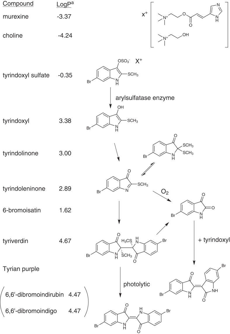
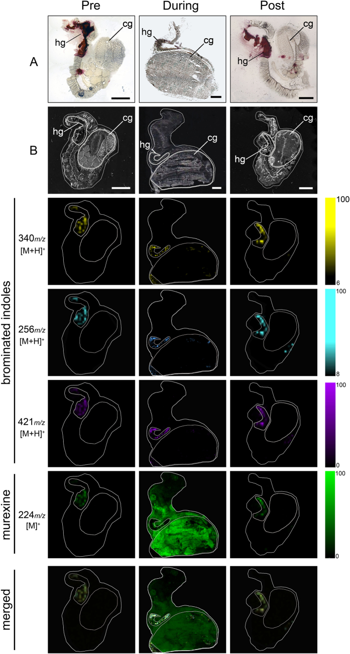
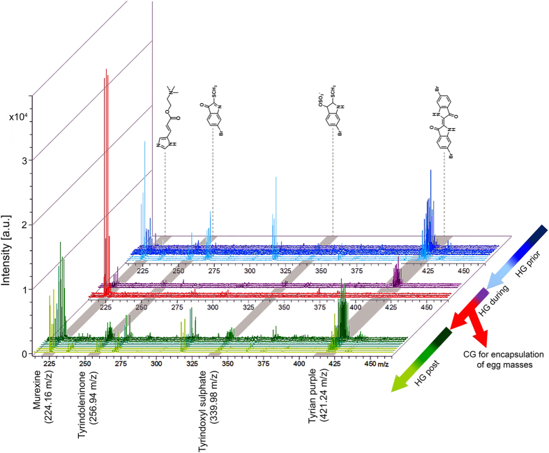
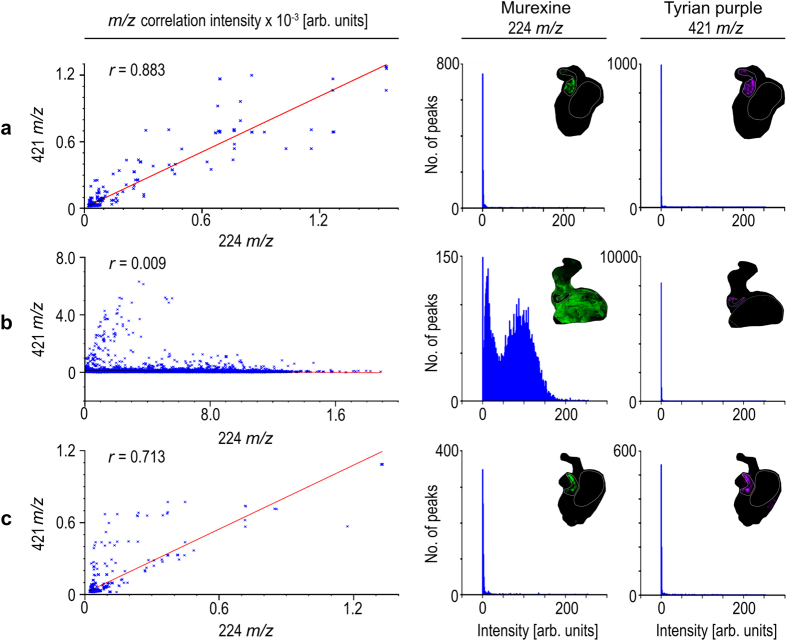
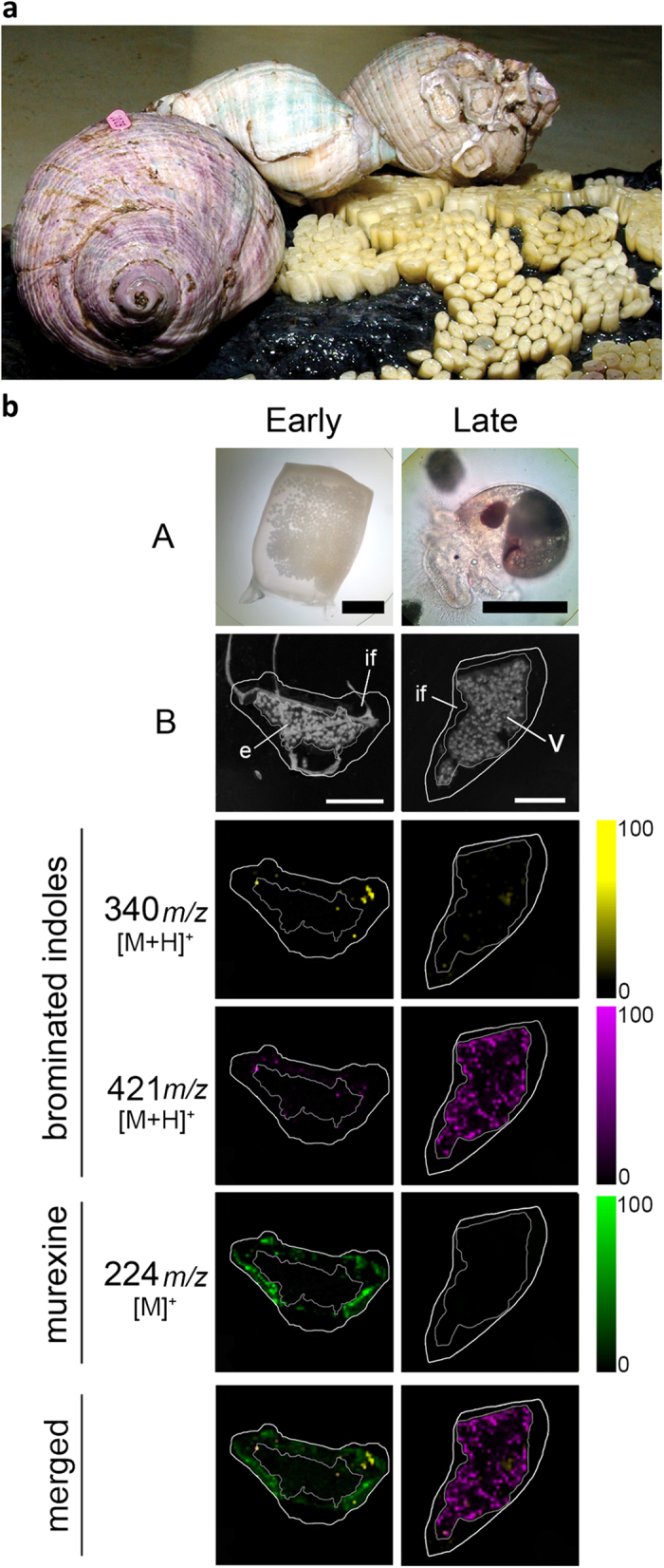
Despite significant advances in chemical ecology, the biodistribution, temporal changes and ecological function of most marine secondary metabolites remain unknown. One such example is the association between choline esters and Tyrian purple precursors in muricid molluscs. Mass spectrometry imaging (MSI) on nano-structured surfaces has emerged as a sophisticated platform for spatial analysis of low molecular mass metabolites in heterogeneous tissues, ideal for low abundant secondary metabolites. Here we applied desorption-ionisation on porous silicon (DIOS) to examine in situ changes in biodistribution over the reproductive cycle. DIOS-MSI showed muscle-relaxing choline ester murexine to co-localise with tyrindoxyl sulfate in the biosynthetic hypobranchial glands. But during egg-laying, murexine was transferred to the capsule gland, and then to the egg capsules, where chemical ripening resulted in Tyrian purple formation. Murexine was found to tranquilise the larvae and may relax the reproductive tract. This study shows that DIOS-MSI is a powerful tool that can provide new insights into marine chemo-ecology.


Figures

Figure 1
Figure 1. The enzymatic, oxidative and photolytic reaction of bioactive compounds found in D. orbita hypobranchial glands (Muricidae, Mollusca) with corresponding solubility indicator.
aLog P was calculated using the chemoinformatics software Molinspiration.
Figure 2
Figure 2. DIOS-MSI maps of secondary metabolites imprinted onto pSi from female D. orbita across the reproductive cycle, in positive ion mode at 100 μm spatial resolution.
(Pre) representative female section sampled 30 days prior to the breeding season. (During) female section sampled during encapsulation. (Post) representative female section sampled 14 days post encapsulation. Maps are compared to (A) histological sections and (B) scanned tissue sections on pSi prior to removal. Tissue regions include (hg) medial hypobranchial gland and (cg) capsule gland. Ion maps m/z 340 corresponds to tyrindoxyl hydrogen sulfate [M+H]+, m/z 256 to tyrindoleninone [M+H]+, m/z 421 to Tyrian purple [M+H]+, and m/z 224 to murexine [M]+. Scale bar set to 2 mm.
Figure 3
Figure 3. D. orbita secondary metabolite mass spectra from across the reproductive cycle.
Spectra show mono- and dibrominated ion clusters for the brominated indoles and murexine detected using DIOS-MSI in positive mode from the medial hypobranchial gland and capsule gland from reproductively active females. (HG) hypobranchial gland; (CG) capsule gland.
Figure 4
Figure 4. Representative correlation plots (left hand panels) for co-localised m/z patterns for murexine against Tyrian purple with corresponding intensity histograms of murexine (middle panels) and Tyrian purple (right hand panels) spot spectra generated in SCiLS Lab imaging software (Bremen, Germany).
Where Tyrian purple and murexine co-localise within; (a) pre-reproductive female tissue section; (b) reproductive female tissue section; and (c) post reproductive female tissue section, with intensities for murexine and Tyrian purple.
Figure 5
Figure 5. D. orbita during egg deposition and DIOS-MSI maps of egg capsules across the developmental period, in positive ion mode at 100 μm spatial resolution.
(a) Reproductive adults during the encapsulation of larvae and early stage capsules adhered to substrate (photo by Rudd, D.). (b) early stage capsule sampled immediately post deposition (left panels A = whole capsule) and late stage capsule after 35 days post deposition (right panels A = encapsulated veliger larvae). DIOS-MSI of the secondary metabolites are compared to (B) scanned cross sections of the egg capsules stamped onto pSi prior to removal. Labels on the imaged regions include; embryo mass (e), intracapsular fluid, (if) and veligar (v) stage larval mass. Ion maps m/z 340 corresponds to tyrindoxyl hydrogen sulfate [M+H]+, m/z 421 to Tyrian purple [M+H]+, and m/z 224 to murexine [M]+. Scale bar set to 1 mm.
Figure 6
Figure 6. The proposed biological role of murexine in the D. orbita egg capsules using a larval motility assay in the presence of 50 ppm murexine extract:
(a) percentage of motile larvae counted using short 30 s videos over 60 min and (b) a video still shot of larvae at time 0 prior to the addition of murexine extract (video online: https://youtu.be/rlCvyyhnXAE).

Similar articles

Cited by

References

    1. Hay M. E. Marine chemical ecology: What’s known and what’s next? J Exp Mar Biol Ecol 200, 103–134 (1996).
    1. Cummins S. F. et al. Characterization of Aplysia enticin and temptin, two novel water-borne protein pheromones that act in concert with attractin to stimulate mate attraction. J Biol Chem 279, 25614–25622 (2004). - PubMed
    1. Cimino G., Passeggio A., Sodano G., Spinella A. & Villani G. Alarm pheromones from the Mediterranean opisthobranch Haminoea navicula. Experientia 47, 61–63 (1991).
    1. Kelley W. P. et al. Characterization of a novel gastropod toxin (6-Bromo-2-mercaptotryptamine) that inhibits shaker K channel activity. J Biol Chem 278, 34934–34942 (2003). - PubMed
    1. Olivera B. M. et al. Diversity of Conus neuropeptides. Science 249, 257–263 (1990). - PubMed

Publication types

MeSH terms

LinkOut - more resources