. 2015 Nov;35(22):3810-28.
doi: 10.1128/MCB.00226-15. Epub 2015 Aug 31.

PPM1G Binds 7SK RNA and Hexim1 To Block P-TEFb Assembly into the 7SK snRNP and Sustain Transcription Elongation

Affiliations

PPM1G Binds 7SK RNA and Hexim1 To Block P-TEFb Assembly into the 7SK snRNP and Sustain Transcription Elongation

Swapna Aravind Gudipaty et al. Mol Cell Biol. 2015 Nov.

Abstract

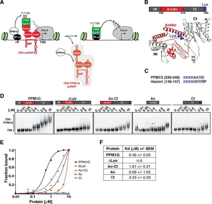
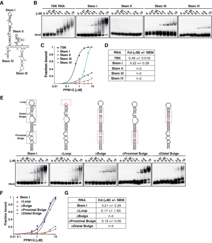
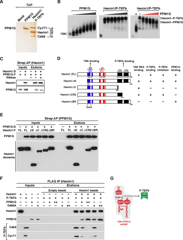
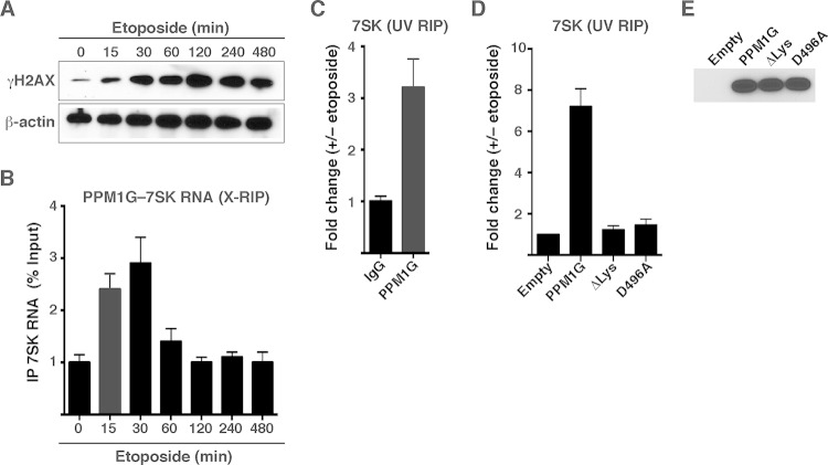
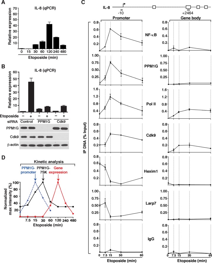
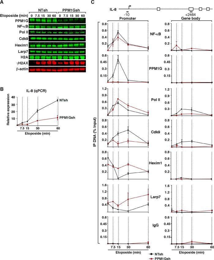
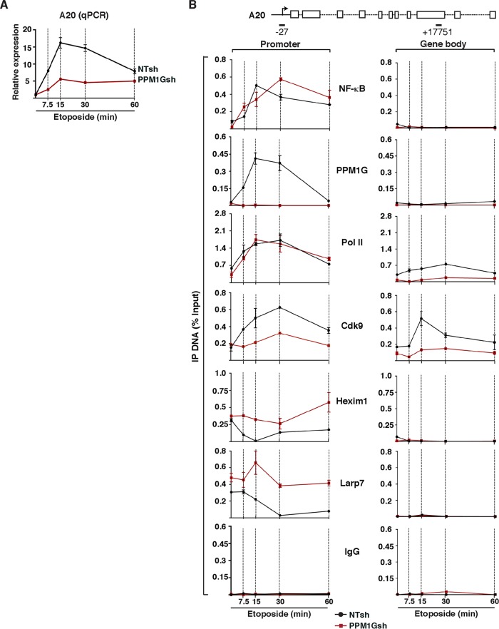
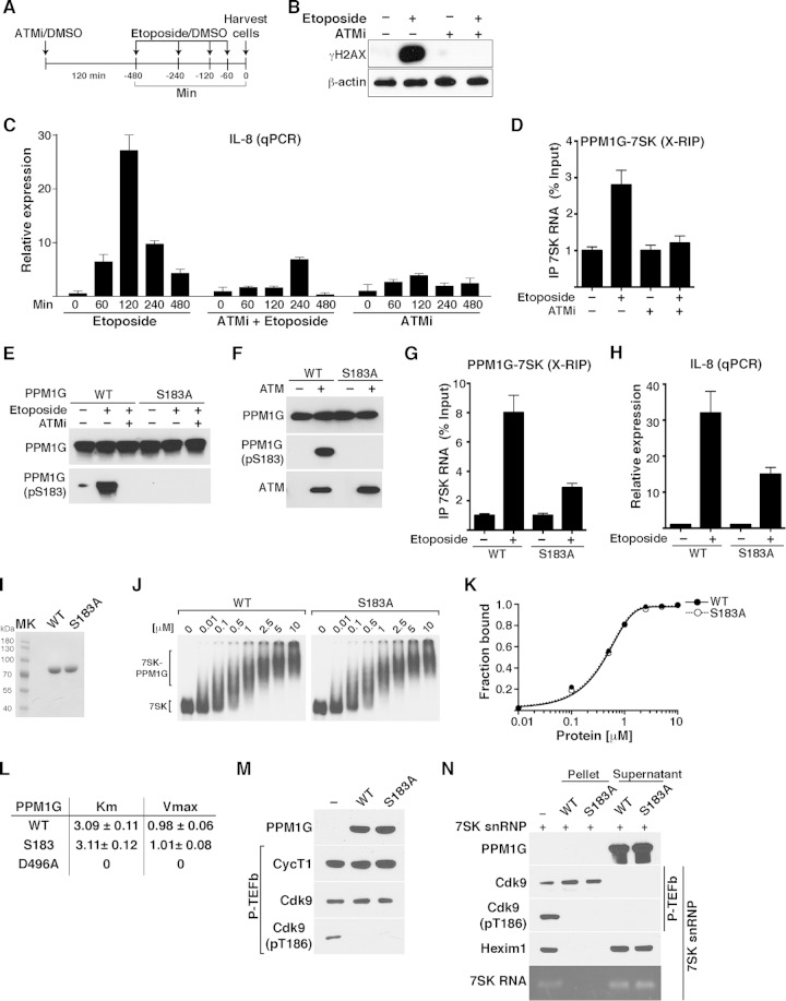
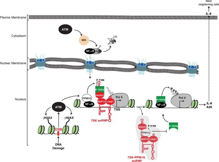
Transcription elongation programs are vital for the precise regulation of several biological processes. One key regulator of such programs is the P-TEFb kinase, which phosphorylates RNA polymerase II (Pol II) once released from the inhibitory 7SK small nuclear ribonucleoprotein (snRNP) complex. Although mechanisms of P-TEFb release from the snRNP are becoming clearer, how P-TEFb remains in the 7SK-unbound state to sustain transcription elongation programs remains unknown. Here we report that the PPM1G phosphatase (inducibly recruited by nuclear factor κB [NF-κB] to target promoters) directly binds 7SK RNA and the kinase inhibitor Hexim1 once P-TEFb has been released from the 7SK snRNP. This dual binding activity of PPM1G blocks P-TEFb reassembly onto the snRNP to sustain NF-κB-mediated Pol II transcription in response to DNA damage. Notably, the PPM1G-7SK RNA interaction is direct, kinetically follows the recruitment of PPM1G to promoters to activate NF-κB transcription, and is reversible, since the complex disassembles before resolution of the program. Strikingly, we found that the ataxia telangiectasia mutated (ATM) kinase regulates the interaction between PPM1G and the 7SK snRNP through site-specific PPM1G phosphorylation. The precise and temporally regulated interaction of a cellular enzyme and a noncoding RNA provides a new paradigm for simultaneously controlling the activation and maintenance of inducible transcription elongation programs.


Figures

FIG 1
FIG 1
The PPM1G phosphatase binds 7SK RNA using its C-terminal Lys-rich domain. (A) Proposed model for the role of PPM1G in the NF-κB transcriptional program. PPM1G is recruited by NF-κB to target genes in response to environmental stimuli, where it dephosphorylates the T-loop of Cdk9 (P-T186), thereby promoting its uncoupling from Hexim1 and its release from the inhibitory, promoter-bound 7SK snRNP complex. In this process, PPM1G subsequently binds 7SK RNA, possibly forming a 7SK-PPM1G snRNP complex lacking P-TEFb. (B) Domain organization of human PPM1G and representation of each domain in the predicted three-dimensional structure. Nt, N-terminal domain; Ac, acidic region; Ct, C-terminal domain; Lys, Lys-rich region. (C) Alignment of PPM1G and Hexim1 Lys-rich regions with their positions indicated in parentheses. (D) Gel shift assays with 7SK RNA and increasing amounts of full-length PPM1G or domains. (E) Binding curves from the gel shifts shown in panel D. (F) Calculations of the apparent dissociation constants (Kdapp) from data in panel E. Kdapp values represent the averages of data from three independent experiments with the standard errors of the means (n = 3). n.d., not determined, because the data set does not enable accurate calculation of the Kdapp values.
FIG 2
FIG 2
PPM1G binds the 7SK RNA stem I distal bulge. (A) Primary sequence and predicted secondary structure of human 7SK RNA with the corresponding stem-loops (Stem). (B) Gel shift assays with full-length PPM1G and 7SK RNA or each of the individual stems. (C) Binding curves from the gel shifts shown in panel B. (D) Calculations of the Kdapp values from data shown in panel C. Kdapp values represent the averages of data from three independent experiments and the standard errors of the means (n = 3). (E) Gel shift assays with PPM1G and WT 7SK stem I or mutants (ΔLoop, ΔBulge, ΔProximal Bulge, and ΔDistal Bulge). The schemes above the gel shifts show the positions of the different RNA elements and the corresponding mutations. (F) Binding curves from the gel shifts shown in panel E. (G) Calculations of the Kdapp from data shown in panel F (means ± standard errors of the means; n = 3). n.d., not determined, because the data set does not enable accurate calculation of the Kdapp values.
FIG 3
FIG 3
PPM1G interacts with Hexim1 and assembles onto 7SK RNA to block P-TEFb binding and 7SK snRNP formation. (A) Affinity purification of Strep-tagged PPM1G and Hexim1–P-TEFb (FLAG-Hexim1, Strep-CycT1, and untagged Cdk9). Proteins were electrophoresed on an SDS-PAGE gel and silver stained. (B) Gel shift assays with full-length 7SK RNA and increasing amounts of PPM1G (left), the Hexim1/P-TEFb complex (middle), and 7SK RNA-bound Hexim1-P-TEFb with increasing amounts of PPM1G (right). (C) Hexim1 binds PPM1G. FLAG-tagged PPM1G (PPM1G:F) was transfected with (+) or without (−) Strep-tagged Hexim1 (Hexim1:S) into HEK 293T cells. Protein lysates treated with (+) or not treated with (−) RNase were used for Strep-Tactin affinity purifications (Strep AP) of Hexim1 and analyzed by Western blotting with the indicated antibodies. (D) Scheme of full-length (FL) Hexim1 and domains alongside their respective biochemical properties in relation to 7SK RNA, P-TEFb, and PPM1G binding (derived from the data shown in panel E). BR denotes the basic region, and AR denotes the acidic region in Hexim1. (E) PPM1G contacts the Hexim1 CR1 domain in vitro. Strep-tagged PPM1G (PPM1G:S) and FLAG-tagged Hexim1 (FL or domains) were purified from HEK 293T cells (inputs). Proteins were used for an in vitro Strep-binding assay where Strep-tagged PPM1G coupled to beads (or empty beads [−]) was incubated with FL Hexim1 or domains in the absence of 7SK RNA, and elutions were analyzed by Western blotting. (F) PPM1G associates with Hexim1 to block P-TEFb binding in vitro. The indicated proteins or protein complexes were bound to beads and incubated with P-TEFb in the presence of 7SK RNA (because it is needed for the Hexim1–P-TEFb interaction) and analyzed by Western blotting. (G) Proposed model where PPM1G associates with Hexim1 on 7SK RNA stem I (referred to as 7SK-PPM1G snRNP) to block Hexim1-mediated P-TEFb recruitment and formation of the 7SK snRNP complex.
FIG 4
FIG 4
PPM1G interacts directly and transiently with 7SK RNA in response to DNA damage. (A) Etoposide induces rapid DNA damage in HeLa cells. Induction of the DNA damage response (as shown by the accumulation of the DNA damage marker γH2AX) in HeLa cells treated with etoposide over a time course is shown. (B) RIP assay (X-RIP) showing the kinetics of the PPM1G-7SK snRNP interaction in response to etoposide treatment. HeLa cells were treated with etoposide as described above for panel A and cross-linked with formaldehyde, and endogenous PPM1G was immunoprecipitated to monitor the interaction between PPM1G and 7SK RNA by qRT-PCR (means ± standard errors of the means; n = 3). (C) UV RIP showing direct interactions between PPM1G and 7SK RNA in response to etoposide treatment. HeLa cells were treated with etoposide (+) or DMSO (−), endogenous PPM1G was immunoprecipitated after UV treatment, and levels of PPM1G-bound 7SK RNA were quantified by qRT-PCR (means ± standard errors of the means; n = 3). (D) The Lys-rich region of catalytically active PPM1G directs the interaction with 7SK RNA in response to etoposide. WT, ΔLys, and catalytically dead D496A mutant PPM1G proteins or an empty vector was transfected into HeLa cells, and cells were subsequently treated with etoposide and UV cross-linked. Levels of coimmunoprecipitating 7SK RNA were quantified by qRT-PCR (means ± standard errors of the means; n = 3). (E) Western blot analyses of lysates of HeLa cells transfected with an empty vector, Strep-tagged PPM1G, and the ΔLys and catalytically dead (D496A) mutants, as described above for panel D.
FIG 5
FIG 5
PPM1G controls transcription of 7SK snRNP-regulated NF-κB target genes in response to DNA damage. (A) Transcription of NF-κB target genes (IL-8 gene) in response to etoposide. HeLa cells were treated with etoposide over a time course, and RNA was isolated for quantitation of gene expression by qRT-PCR. The expression level of IL-8 was normalized to that of β-actin (means ± standard errors of the means; n = 3). (B) KD of PPM1G and Cdk9 abolishes transcription of the IL-8 gene in response to etoposide. HeLa cells were transfected with nontarget control, PPM1G, or Cdk9 siRNAs for 48 h to knock down the indicated proteins, followed by treatment with etoposide (+) or DMSO (−) for 120 min. Total RNA was extracted, and IL-8 gene expression was quantified by qRT-PCR (means ± standard errors of the means; n = 3). The bottom panel shows Western blotting for validation of the RNAi-mediated KD. (C) ChIP assay showing levels of NF-κB, PPM1G, Pol II, and 7SK snRNP components (Cdk9, Hexim1, and Larp7) at the IL-8 promoter and gene body in response to a time course of etoposide treatment (means ± standard errors of the means; n = 3). Normal serum (IgG) was used as a negative control to demonstrate the specificity of enrichment with the antibodies used. (D) Kinetic plots for IL-8 gene expression (as determined by qRT-PCR as described above for panel A), PPM1G recruitment to the IL-8 promoter (as determined by ChIP as described above for panel C), and PPM1G-7SK RNA interactions (as determined by an X-RIP assay as described for Fig. 4B) in HeLa cells in response to etoposide. Note that the time scale (x axis) is on a log2 scale for better data visualization.
FIG 6
FIG 6
PPM1G functions downstream of NF-κB and Pol II recruitment to gene promoters to induce 7SK snRNP disassembly and Pol II elongation in response to DNA damage. (A) HeLa cells were transduced with lentiviruses (pLKO) inducibly expressing nontarget (NTsh) or PPM1G (PPM1Gsh) shRNAs, induced with IPTG for 3 days, and treated with etoposide. PPM1G KD efficiency, induction of DNA damage (γH2AX), and stability of transcriptional components were evaluated by Western blotting over time. (B) PPM1G KD antagonizes IL-8 gene expression in response to etoposide. The cell lines from panel A were used for total RNA extraction, and IL-8 gene expression in response to etoposide was quantified by qRT-PCR (means ± standard errors of the means; n = 3). The data were normalized to levels of the β-actin transcript. (C) ChIP assay of cell lines from panel A showing levels of NF-κB, PPM1G, Pol II, and 7SK snRNP components (Cdk9, Hexim1, and Larp7) at the IL-8 promoter-proximal region (−10 amplicon) and gene body (+2464 amplicon) in response to a time course of etoposide treatment (means ± standard errors of the means; n = 3). Normal serum (IgG) was used as a negative control to demonstrate the specificity of enrichment with the antibodies used.
FIG 7
FIG 7
PPM1G is required for NF-κB-mediated Pol II transcription elongation at the A20 gene in response to etoposide. (A) PPM1G KD abolishes A20 gene expression in response to etoposide. The cell lines from Fig. 6A (HeLa NTsh and PPM1Gsh) were used for total RNA extraction, and A20 gene expression in response to etoposide was quantified by qRT-PCR (means ± standard errors of the means; n = 3). The data were normalized to levels of the β-actin transcript. (B) ChIP assay of cell lines from Fig. 6A showing levels of NF-κB, PPM1G, Pol II, and 7SK snRNP components (Cdk9, Hexim1, and Larp7) at the A20 promoter-proximal region (−27 amplicon) and gene body (+17751 amplicon) in response to a time course of etoposide treatment (means ± standard errors of the means; n = 3). Normal serum (IgG) was used as a negative control to demonstrate the specificity of enrichment with the antibodies used.
FIG 8
FIG 8
PPM1G-7SK RNA interaction and activation of NF-κB transcription in response to DNA damage are ATM kinase dependent. (A) Scheme showing the protocol used to pretreat HeLa cells with ATMi (or DMSO as a control) for 120 min, followed by a time course of etoposide treatment or DMSO. (B) Validation of ATM inhibition with an ATMi by Western blotting with γH2AX antibody. HeLa cells were pretreated for 120 min with an ATMi or DMSO as a control, followed by a 120-min etoposide or DMSO treatment. Cell pellets were used for Western blotting with the indicated antibodies (β-actin was used as a loading control). (C) ATM inhibition blocks NF-κB transcription (IL-8 gene) from HeLa cells in response to etoposide. Cells were pretreated with an ATMi or vehicle (DMSO) for 120 min and sequentially treated with etoposide or vehicle (DMSO) for the time points indicated. Total RNA was then extracted, and the IL-8 gene expression level, normalized to the β-actin level, was calculated by qRT-PCR (means ± standard errors of the means; n = 3). (D) ATM inhibition abolishes the PPM1G-7SK protein-RNA interaction in response to etoposide. HeLa cells were pretreated (+) or not pretreated (−) with an ATMi (120 min), followed by a 30-min treatment with etoposide or DMSO (−). Cells were cross-linked with formaldehyde, and endogenous PPM1G was immunoprecipitated to quantify the levels of coprecipitated 7SK RNA in response to etoposide by qRT-PCR (means ± standard errors of the means; n = 3). (E) PPM1G is phosphorylated at Ser183 (S183) in response to etoposide, and an ATMi blocks site-specific PPM1G phosphorylation (pS183) upon etoposide treatment. HeLa cells were transfected with Strep-tagged WT PPM1G or the S183A mutant and treated as described above for panel D. Proteins were affinity purified and used for Western blot assays with the indicated antibodies. (F) PPM1G is phosphorylated by the ATM kinase at S183. Strep-affinity-purified WT PPM1G or the S183A mutant was incubated with (+) or without (−) ATM kinase, and Western blot analyses (total PPM1G and the pS183 form) were performed. (G) Mutation of the Ser183 residue (S183A) in PPM1G reduces the PPM1G-7SK RNA interaction in response to etoposide. HeLa cells were transfected with Strep-tagged WT PPM1G or the S183A mutant, treated with etoposide (+) or DMSO (−) for 30 min, and cross-linked with formaldehyde, and a RIP assay (X-RIP) was performed to quantitate the association between PPM1G and 7SK RNA by using qRT-PCR (means ± standard errors of the means; n = 3). (H) Mutation of Ser183 in PPM1G (S183A) reduces activation of IL-8 gene expression in response to etoposide. HeLa cells were transfected with siRNA-resistant (HS_PPM1G_6 [see Table S3 at http://www.utsouthwestern.edu/labs/dorso/research/lab-projects.html]) Strep-tagged WT PPM1G or the S183A mutant and retransfected 24 h later with a PPM1G siRNA to knock down endogenous PPM1G. Forty-eight hours later (at which time the PPM1G KD level was ∼80%), cells were treated with etoposide (+) or DMSO (−) for 120 min, RNA was extracted, and IL-8 gene expression was quantified by qRT-PCR and normalized to the β-actin level (means ± standard errors of the means; n = 3). (I) Strep-tagged WT PPM1G and S183A mutant proteins were affinity purified and visualized by Coomassie staining. MK, protein molecular size marker. (J) Gel shift assays with 7SK RNA and increasing amounts of WT PPM1G or the S183A mutant. (K) Binding curves for the gel shifts shown in panel J. (L) Determination of the kinetic parameters Km and Vmax for WT PPM1G, the S183A mutant, and the catalytically dead mutant (D496A) on the phosphatase substrate pNPP. (M) Purified P-TEFb was incubated with WT PPM1G or the S183A mutant under dephosphorylation conditions, and Cdk9 T-loop phosphorylation at Thr186 (pT186) was monitored by Western blotting. (N) The S183A mutation in PPM1G does not affect the enzymatic release of P-TEFb from the 7SK snRNP complex. 7SK-bound P-TEFb complexes were incubated with WT PPM1G or the S183A mutant under dephosphorylation conditions. Subsequent purification of P-TEFb using FLAG beads was done to monitor released (supernatant) and retained (pellet) components by Western blotting.
FIG 9
FIG 9
Model depicting the functional interplay between PPM1G, the 7SK snRNP, and ATM kinase during the activation of the NF-κB transcriptional program in response to DNA damage. In response to double-strand breaks (DSB), ATM activates the NF-κB signaling pathway through phosphorylation of the NEMO subunit of the IKK complex, which in turn phosphorylates the NF-κB inhibitor (IκB), leading to its ubiquitination (Ub) and proteasomal degradation. NF-κB translocates to the nucleus, where it recruits ATM-phosphorylated PPM1G to target genes such as the IL-8 gene, dephosphorylating and releasing the P-TEFb kinase from the promoter-bound 7SK snRNP complex. Upon its release, P-TEFb phosphorylates (P) paused Pol II in proximity to the transcription start site (TSS) to promote transcriptional pause release. After releasing P-TEFb, the inhibitory snRNP subunits are evicted from chromatin, and phosphorylated PPM1G binds 7SK RNA along with Hexim1 to prevent the reassociation of P-TEFb back into the snRNP to sustain transcription elongation. Once the damage is resolved, this regulatory circuitry subsides, PPM1G is dislodged from the snRNP, and P-TEFb is recycled back to promote the formation of the inhibitory 7SK snRNP at the promoter, thereby blocking Pol II pause release.

Similar articles

Cited by

References

    1. Smale ST. 2010. Selective transcription in response to an inflammatory stimulus. Cell 140:833–844. doi:10.1016/j.cell.2010.01.037. - DOI - PMC - PubMed
    1. Adelman K, Lis JT. 2012. Promoter-proximal pausing of RNA polymerase II: emerging roles in metazoans. Nat Rev Genet 13:720–731. doi:10.1038/nrg3293. - DOI - PMC - PubMed
    1. Smith E, Shilatifard A. 2013. Transcriptional elongation checkpoint control in development and disease. Genes Dev 27:1079–1088. doi:10.1101/gad.215137.113. - DOI - PMC - PubMed
    1. Jonkers I, Lis JT. 2015. Getting up to speed with transcription elongation by RNA polymerase II. Nat Rev Mol Cell Biol 16:167–177. doi:10.1038/nrm3953. - DOI - PMC - PubMed
    1. Buratowski S. 2009. Progression through the RNA polymerase II CTD cycle. Mol Cell 36:541–546. doi:10.1016/j.molcel.2009.10.019. - DOI - PMC - PubMed

Publication types

MeSH terms