

Border Cell Migration: A Model System for Live Imaging and Genetic Analysis of Collective Cell Movement
- PMID: 26324431
- PMCID: PMC4762686
- DOI: 10.1007/978-1-4939-2851-4_6
Border Cell Migration: A Model System for Live Imaging and Genetic Analysis of Collective Cell Movement
Abstract
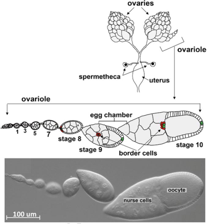
Border cell migration in the Drosophila ovary has emerged as a genetically tractable model for studying collective cell movement. Over many years border cell migration was exclusively studied in fixed samples due to the inability to culture stage 9 egg chambers in vitro. Although culturing late-stage egg chambers was long feasible, stage 9 egg chambers survived only briefly outside the female body. We identified culture conditions that support stage 9 egg chamber development and sustain complete migration of border cells ex vivo. This protocol enables one to compare the dynamics of egg chamber development in wild-type and mutant egg chambers using time-lapse microscopy and taking advantage of a multiposition microscope with a motorized imaging stage. In addition, this protocol has been successfully used in combination with fluorescence resonance energy transfer biosensors, photo-activatable proteins, and pharmacological agents and can be used with wide-field or confocal microscopes in either an upright or an inverted configuration.
Figures
References
-
- Ilina O, Friedl P. Mechanisms of collective cell migration at a glance. J Cell Sci. 2009;122:3203–3208. - PubMed
-
- Aman A, Piotrowski T. Cell migration during morphogenesis. Dev Biol. 2010;341:20–33. - PubMed
-
- Weijer CJ. Collective cell migration in development. J Cell Sci. 2009;122:3215–3223. - PubMed
-
- Lopez-Schier H. Fly fishing for collective cell migration. Curr Opin Genet Dev. 2010;20:428–432. - PubMed
-
- Rorth P. Collective cell migration. Annu Rev Cell Dev Biol. 2009;25:407–429. - PubMed
Publication types
MeSH terms
Grants and funding
LinkOut - more resources
Full Text Sources
Other Literature Sources
Molecular Biology Databases
