. 2015 Nov;197(22):3533-44.
doi: 10.1128/JB.00606-15. Epub 2015 Aug 31.

NagR Differentially Regulates the Expression of the glmS and nagAB Genes Required for Amino Sugar Metabolism by Streptococcus mutans

Affiliations

NagR Differentially Regulates the Expression of the glmS and nagAB Genes Required for Amino Sugar Metabolism by Streptococcus mutans

Lin Zeng et al. J Bacteriol. 2015 Nov.

Abstract

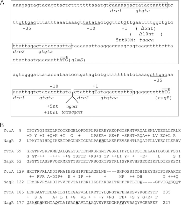
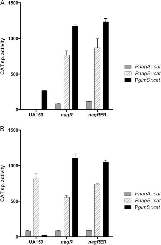
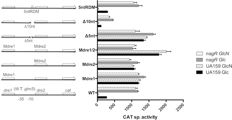
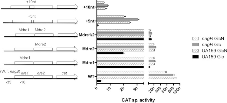
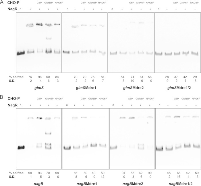
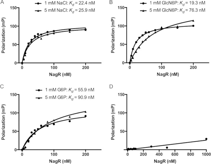
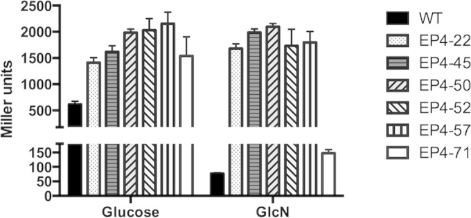
The ability of bacteria to metabolize glucosamine (GlcN) and N-acetyl-d-glucosamine (GlcNAc) is considered important for persistent colonization of the oral cavity. In the dental caries pathogen Streptococcus mutans, the NagR protein regulates the expression of glmS, which encodes a GlcN-6-P synthetase, and nagA (GlcNAc-6-P deacetylase) and nagB (GlcN-6-P deaminase), which are required for the catabolism of GlcNAc and GlcN. Two NagR-binding sites (dre) were identified in each of the promoter regions for nagB and glmS. Using promoter-reporter gene fusions, the role of each dre site was examined in the regulation of glmS and nagB promoter activities in cells grown with glucose, GlcNAc, or GlcN. A synergistic relationship between the two dre sites in the glmS promoter that required proper spacing was observed, but that was not the case for nagB. Binding of purified NagR to DNA fragments from both promoter regions, as well as to dre sites alone, was strongly influenced by particular sugar phosphates. Using a random mutagenesis approach that targeted the effector-binding domain of NagR, mutants that displayed aberrant regulation of both the glmS and nagAB genes were identified. Collectively, these findings provide evidence that NagR is essential for regulation of genes for both the synthesis and catabolism of GlcN and GlcNAc in S. mutans, and that NagR engages differently with the target promoter regions in response to specific metabolites interacting with the effector-binding domain of NagR.

Importance: Glucosamine and N-acetylglucosamine are among the most abundant naturally occurring sugars on the planet, and they are catabolized by many bacterial species as sources of carbon and nitrogen. Representing a group called lactic acid bacteria (LAB), the human dental caries pathogen Streptococcus mutans is shown to differ from known paradigm organisms in that it possesses a GntR/HutC-type regulator, NagR, that is required for the regulation of both catabolism of GlcN and biosynthesis. Results reported here reveal a simple and elegant mechanism whereby NagR differentially regulates two opposing biological processes by surveying metabolic intermediates. This study provides insights that may contribute to the development of novel therapeutic tools to combat dental caries and other infectious diseases.


Figures

FIG 1
FIG 1
Schematic of the upstream regions of the glmS and nagB genes (A) and alignment of sequences of NagR from S. mutans and YvoA/NagR from B. subtilis (B). (A) The locations of the predicted promoter elements are underlined and labeled as −35, −10, and +1 (transcription initiation site), and the start codon of each gene is represented by an arrow. The putative binding sites (dre1 and dre2) (see Table S2 in the supplemental material for the consensus sequence) for NagR are shown in boxes and labeled, with engineered modifications described below in italics. Also delineated are deletions and insertions introduced to alter the phasing of the dre sites. (B) Underlined are sites of mutations engineered in the DNA-binding domain (aa 2 to 69) near the N terminus of NagR and random mutations isolated in the putative effector-binding domain (aa 88 to 226) near the C terminus.
FIG 2
FIG 2
CAT assays measuring the promoter activities of glmS, nagB, and nagA, with each fused to a promoterless cat gene, in the backgrounds of wild-type UA159, a nagR null mutant, and the nagR-ER (E30A, R31A) mutant. Bacterial strains were cultivated in TV medium supplemented with glucose (A) or GlcN (B) as sole carbohydrates and harvested and processed for CAT assays as detailed in Materials and Methods. sp., specific. The bars represent the average activities (expressed in nanomoles per milligram of protein per minute) of three independent cultures, and the error bars denote standard deviations.
FIG 3
FIG 3
CAT specific activities (expressed in nanomoles of chloramphenicol acetylated per milligram of protein per minute) of fusions constructed using wild-type (W.T.) and various mutant versions of the glmS promoter in the backgrounds of UA159 and a nagR null mutant. Mutations in Mdre1, Mdre2, or Mdre1/2 are replacements of a conserved TATAC sequence in corresponding dre sites with GTGTA; Δ5nt and Δ10nt are truncations of a 5- or 10-nt sequence located between two dre sites without compromising the −10 promoter structure; 5ntRDM is a result of sequence randomization of the same 5 nucleotides in Δ5nt. Bacteria were cultured in TV base medium supplemented with 0.5% glucose (Glc) or GlcN and then harvested and processed for CAT assays as detailed in Materials and Methods. Results are derived from triplicate assays from three independent cultures and are presented as averages ± standard deviations.
FIG 4
FIG 4
CAT specific activities (expressed in nanomoles per milligram of protein per minute) of fusions constructed with wild-type (W.T.) and mutant versions of the nagB promoter region in the background of UA159 and its otherwise-isogenic nagR null mutant. Mutation in Mdre1 is a replacement of a conserved TATAC sequence in dre1 with GTGTA, while Mdre2 is a replacement of similarly conserved TAGACC in dre2 with GTGTAA. +5nt and +10nt are insertions of a 5- and 10-nt random sequence, respectively, between two dre sites. Bacteria were cultured in TV base medium supplemented with 0.5% glucose (Glc) or GlcN and then harvested for CAT assays as detailed in Materials and Methods. Results are derived from three independent cultures assayed in triplicate and are presented as averages ± standard deviations.
FIG 5
FIG 5
Electrophoretic mobility shift assay (EMSA). One femtomole of biotinylated DNA probe containing regions upstream of the glmS (A) or nagB (B) coding sequences, including the respective dre mutants, was used in each reaction with 0, 1.3 μM (A), or 0.3 μM (B) NagR protein. When indicated, 20 mM glucose-6-P (G6P), GlcN-6-P (GlcN6P), or GlcNAc-6-P (NAG6P) also was added to the reaction mixture. Indicated below each lane are the average percentages and standard deviations (S.D.) of probe being shifted relative to the control lane of the same gel, calculated using the results from two independent repeats.
FIG 6
FIG 6
NagR binds specifically to a DNA probe containing a consensus dre sequence. Fluorescence polarization (FP) assays were performed using a fluorescein-labeled dre probe and purified NagR protein in the presence of additional NaCl (A), GlcN-6-P (B), or G-6-P (C). (D) As a negative control, a similarly labeled DNA oligonucleotide containing a known catabolite response element (cre) also was used in binding with NagR.
FIG 7
FIG 7
β-Galactosidase activities of the wild type (WT) and various nagR point mutants (EP4-22 to EP4-71) carrying the PglmS::lacZ fusion. Strains were grown to the exponential phase (OD600 of ≈0.5) in TV-glucose or TV-GlcN medium before being harvested, permeabilized by treatment with toluene-acetone (1:9, vol/vol), and subjected to Miller assays. Results here each represent the averages and standard deviations (error bars) from three biological repeats.

Similar articles

Cited by

References

    1. Postma PW, Lengeler JW, Jacobson GR. 1993. Phosphoenolpyruvate:carbohydrate phosphotransferase systems of bacteria. Microbiol Rev 57:543–594. - PMC - PubMed
    1. Abranches J, Chen YY, Burne RA. 2003. Characterization of Streptococcus mutans strains deficient in EIIABMan of the sugar phosphotransferase system. Appl Environ Microbiol 69:4760–4769. doi:10.1128/AEM.69.8.4760-4769.2003. - DOI - PMC - PubMed
    1. Moye ZD, Burne RA, Zeng L. 2014. Uptake and metabolism of N-acetylglucosamine and glucosamine by Streptococcus mutans. Appl Environ Microbiol 80:5053–5067. doi:10.1128/AEM.00820-14. - DOI - PMC - PubMed
    1. Zeng L, Das S, Burne RA. 2010. Utilization of lactose and galactose by Streptococcus mutans: transport, toxicity, and carbon catabolite repression. J Bacteriol 192:2434–2444. doi:10.1128/JB.01624-09. - DOI - PMC - PubMed
    1. Plumbridge JA. 1991. Repression and induction of the nag regulon of Escherichia coli K-12: the roles of nagC and nagA in maintenance of the uninduced state. Mol Microbiol 5:2053–2062. doi:10.1111/j.1365-2958.1991.tb00828.x. - DOI - PubMed

Publication types

MeSH terms

Substances