. 2015 Nov;197(22):3563-72.
doi: 10.1128/JB.00603-15. Epub 2015 Aug 31.

A Matter of Timing: Contrasting Effects of Hydrogen Sulfide on Oxidative Stress Response in Shewanella oneidensis

Affiliations

A Matter of Timing: Contrasting Effects of Hydrogen Sulfide on Oxidative Stress Response in Shewanella oneidensis

Genfu Wu et al. J Bacteriol. 2015 Nov.

Abstract

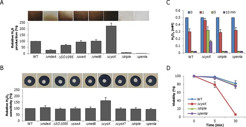
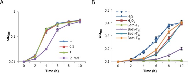
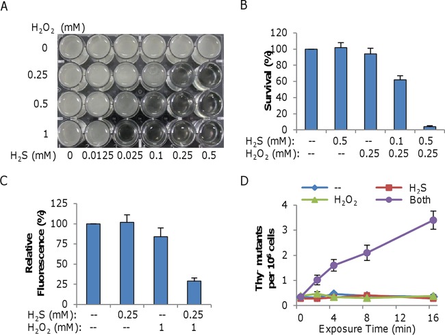
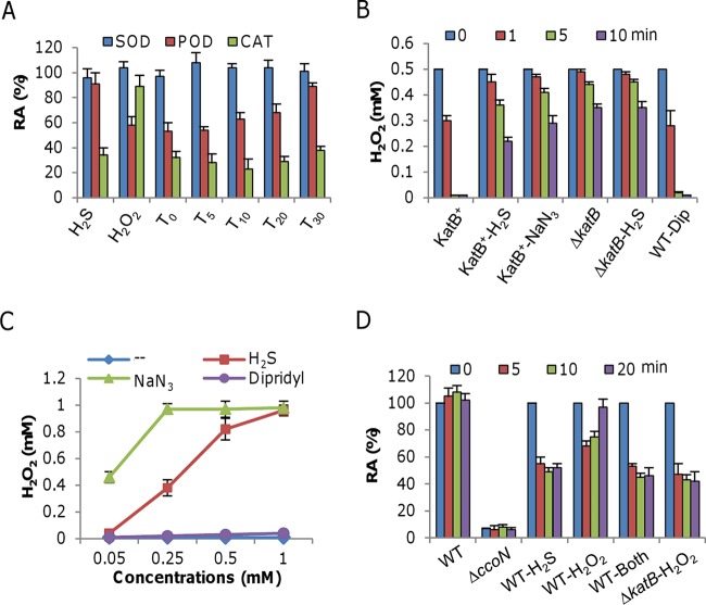
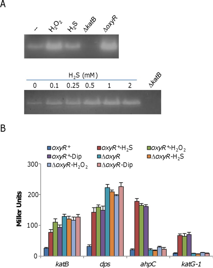
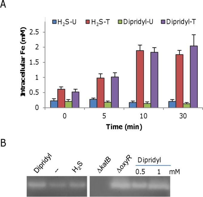
Hydrogen sulfide (H2S), well known for its toxic properties, has recently become a research focus in bacteria, in part because it has been found to prevent oxidative stress caused by treatment with some antibiotics. H2S has the ability to scavenge reactive oxygen species (ROS), thus preventing oxidative stress, but it is also toxic, leading to conflicting reports of its effects in different organisms. Here, with Shewanella oneidensis as a model, we report that the effects of H2S on the response to oxidative stress are time dependent. When added simultaneously with H2O2, H2S promoted H2O2 toxicity by inactivating catalase, KatB, a heme-containing enzyme involved in H2O2 degradation. Such an inhibitory effect may apply to other heme-containing proteins, such as cytochrome cbb3 oxidase. When H2O2 was supplied 20 min or later after the addition of H2S, the oxidative-stress-responding regulator OxyR was activated, resulting in increased resistance to H2O2. The activation of OxyR was likely triggered by the influx of iron, a response to lowered intracellular iron due to the iron-sequestering property of H2S. Given that Shewanella bacteria thrive in redox-stratified environments that have abundant sulfur and iron species, our results imply that H2S is more important for bacterial survival in such environmental niches than previously believed.

Importance: Previous studies have demonstrated that H2S is either detrimental or beneficial to bacterial cells. While it can act as a growth-inhibiting molecule by damaging DNA and denaturing proteins, it helps cells to combat oxidative stress. Here we report that H2S indeed has these contrasting biological functions and that its effects are time dependent. Immediately after H2S treatment, there is growth inhibition due to damage of heme-containing proteins, at least to catalase and cytochrome c oxidase. In contrast, when added a certain time later, H2S confers an enhanced ability to combat oxidative stress by activating the H2O2-responding regulator OxyR. Our data reconcile conflicting observations about the functions of H2S.


Figures

FIG 1
FIG 1
Endogenous H2S does not protect bacteria against H2O2. (A) H2S production in various S. oneidensis strains. Lead acetate-soaked paper strips show a brown or black PbS stain as a result of reaction with H2S under aerobic conditions. The relative H2S levels shown were obtained by normalization to the average level of the wild type (WT), which was set to 100%. The ΔtriplemdeA ΔSO1095 ΔsseA) mutant strain lacks the ability to produce H2S via cysteine metabolism, while the Δpenta mutant strain carries additional deletions in the psrA and sirA genes, thus removing the ability to produce H2S through respiration. (B) H2O2 sensitivity assay. Paper disks of 6 mm loaded with 10 μl of 5 M H2O2 were placed on a bacterial lawn and photographed after 16 h at 30°C. The relative H2O2 susceptibilities shown were obtained by normalization to the average level of the wild type, which was set to 100%. The ΔcysKC mutant is a complemented mutant carrying a copy of the cysK gene integrated into the chromosome and produces H2S like the wild type. (C) H2O2 consumption assay. H2O2 at 0.5 mM was added to mid-log-phase cultures (OD600 of ∼0.2, the same afterward), and the H2O2 remaining at the time points indicated was measured. (D) Survival assay. One millimolar H2O2 was added to mid-log-phase cultures. After 5 and 30 min, samples were diluted and plated on LB. Colony counting was done after 24 h. For panels A and B, experiments were performed five times and representative results are shown. In panels B and D, data are reported as the mean ± SD (n = 4).
FIG 2
FIG 2
Effects of H2S, H2O2, and both together on the growth of the S. oneidensis Δpenta mutant. Mid-log-phase cultures were inoculated into LB medium containing the chemicals indicated and incubated statically at 30°C in 24-well plates. (A) Effect of H2S (from NaHS) on growth. (B) Effects of 0.1 mM H2S, 1 mM H2O2, and both on the growth of the Δpenta mutant strain. T0, T10, T20, and T30 represent the addition of H2O2 at the same time as H2S and 10, 20, and 30 min later, respectively. In both panels A and B, -- represents the LB control. Data are reported as the mean ± SD (n = 4).
FIG 3
FIG 3
H2S promotes H2O2 killing when added promptly after H2O2. (A) Synergistic inhibition of S. oneidensis growth by H2S and H2O2 when added at the same time. Cells at an OD600 of ∼0.01 were inoculated into LB in 24-well plates containing H2S, H2O2, or both at the concentrations indicated and incubated statically at 30°C. The plates were photographed 24 h after inoculation. Experiments were performed five times, and similar results were obtained. (B) Mid-log-phase cells (OD600 of ∼0.2) were treated for 20 min with H2S, H2O2, or both at the concentrations indicated. The treated cultures were diluted, plated on LB, and incubated at 30°C. Survival was calculated as the ratio of the number of colonies in the treated cultures to that in the untreated control. Only plates containing 100 to 300 colonies were counted. (C) H2S-H2O2 treatment generates extensive DNA damage, as determined by qPCR. Mid-log-phase cells were treated with 0.25 mM H2S, 1.0 mM H2O2, or both. Total genomic DNA was extracted, and qPCR was performed with equivalent amounts of template DNA. The relative fluorescence was normalized to that of the untreated control. Data are reported as the mean ± SD (n = 4). (D) H2S-H2O2 treatment generates extensive DNA damage, as determined by thyA mutation analysis. Mid-log-phase cells grown in LB were diluted 100-fold with fresh LB (--) or with LB containing 0.1 mM H2S, 0.25 mM H2O2, or both. At each time point, CAT was added to degrade H2O2, and both total viability and the frequency of thyA mutants were determined. Data are reported as the mean ± SD (n = 4).
FIG 4
FIG 4
Effects of H2S, H2O2, and both on selected heme-containing enzymes. (A) Mid-log-phase cells were harvested, washed, and sonicated for the preparation of crude enzyme extracts. H2S at 0.25 mM, H2O2 at 0.5 mM, or the two together were added to crude enzyme extracts at the times indicated, and SOD, POD, and CAT activities were measured. Subscript numbers are the times (in minutes) when H2O2 was added after the addition of H2S. The activities of the treated samples were normalized to the activity of the untreated control and are reported as relative activities (RA). Importantly, the activities of all three enzymes from untreated samples were found to be stable for 30 min. (B) Crude enzyme extracts prepared as described for panel A were treated with the reagents indicated and assayed for H2O2 degradation. Concentrations: H2S, 0.25 mM; NaN3, 0.1 mM; dipyridyl (Dip), 0.5 mM. WT, wild type. (C) Two thousand five hundred units of bovine liver CAT was treated with the chemicals indicated at various concentrations for 5 min and then assayed for the ability to degrade 1 mM H2O2 at 37°C compared to the control (--). After 5 min, the remaining H2O2 concentrations were measured. (D) Cco assay. Crude enzyme extracts prepared as described for panel A were treated with the reagents indicated. The ΔccoN mutant strain, lacking the essential catalytic subunit of the cbb3 oxidase, was used as a negative control, and the ΔkatB mutant strain was also included for reference. All chemicals were added at the same time. Concentrations: H2S, 0.25 mM; H2O2, 0.5 mM. In all panels, data are reported as the mean ± SD (n = 4).
FIG 5
FIG 5
H2S induces the OxyR-mediated stress response. (A) CAT staining analysis. Cells were harvested just prior to (−) and 30 min after the addition of 0.25 mM H2S or 0.1 mM H2O2 (top). Protein samples of ∼10 μg from the cell lysates indicated were separated by native PAGE and stained for CAT activity. The ΔkatB and ΔoxyR mutant strains (constitutive high-level expression) were used as negative and positive controls. In the analysis shown at the bottom, various concentrations of H2S were examined for the ability to induce expression of the katB gene. (B) Impact of H2S on the expression of four members of the OxyR regulon. β-Galactosidase assays were carried out with lacZ reporter vectors. Cells grown to mid-log phase were treated with the chemicals indicated for 30 min and then harvested for the assays. Concentrations: H2S, 0.25 mM; H2O2, 0.2 mM; dipyridyl (Dip), 0.25 mM. β-Galactosidase activities are reported as the mean ± SD (n = 4). Similar results were obtained by qRT-PCR assay (see Fig. S3 in the supplemental material).
FIG 6
FIG 6
H2S induces the OxyR-mediated stress response. (A) Intracellular iron levels induced by H2S. One-liter cultures grown to mid-log phase (OD600 of ∼0.2) were harvested just before (0 min) and 5, 10, and 30 min after the addition of 0.25 mM H2S or 0.2 mM dipyridyl, and the unincorporated (U) and total (T) intracellular iron concentrations were measured. The experiments were performed at least three times. Error bars show standard deviations. (B) CAT staining analysis. Cells were harvested 30 min after the addition of 0.25 mM H2S or 0.2 mM dipyridyl along with the untreated control (--). Ten-microgram samples of protein from the cell lysates indicated were separated by native PAGE and stained for CAT activity. The ΔkatB and ΔoxyR mutant strains (constitutive high-level expression) were used as negative and positive controls. In the right panel, two higher concentrations of dipyridyl were tested for enhanced induction of the katB gene. The data are representative of three independent experiments.

References

    1. Wang R. 2012. Physiological implications of hydrogen sulfide: a whiff exploration that blossomed. Physiol Rev 92:791–896. doi: 10.1152/physrev.00017.2011. - DOI - PubMed
    1. Kimura H. 2014. Production and physiological effects of hydrogen sulfide. Antioxid Redox Signal 20:783–793. doi: 10.1089/ars.2013.5309. - DOI - PMC - PubMed
    1. Shatalin K, Shatalina E, Mironov A, Nudler E. 2011. H2S: a universal defense against antibiotics in bacteria. Science 334:986–990. doi: 10.1126/science.1209855. - DOI - PubMed
    1. Eghbal MA, Pennefather PS, O'Brien PJ. 2004. H2S cytotoxicity mechanism involves reactive oxygen species formation and mitochondrial depolarisation. Toxicology 203:69–76. doi: 10.1016/j.tox.2004.05.020. - DOI - PubMed
    1. Lalucat J, Bennasar A, Bosch R, García-Valdés E, Palleroni NJ. 2006. Biology of Pseudomonas stutzeri. Microbiol Mol Biol Rev 70:510–547. doi: 10.1128/MMBR.00047-05. - DOI - PMC - PubMed

Publication types

MeSH terms