. 2015 Sep;146(3):221-32.
doi: 10.1085/jgp.201411350.

Integrins mediate mechanical compression-induced endothelium-dependent vasodilation through endothelial nitric oxide pathway

Affiliations

Integrins mediate mechanical compression-induced endothelium-dependent vasodilation through endothelial nitric oxide pathway

Xiao Lu et al. J Gen Physiol. 2015 Sep.

Abstract

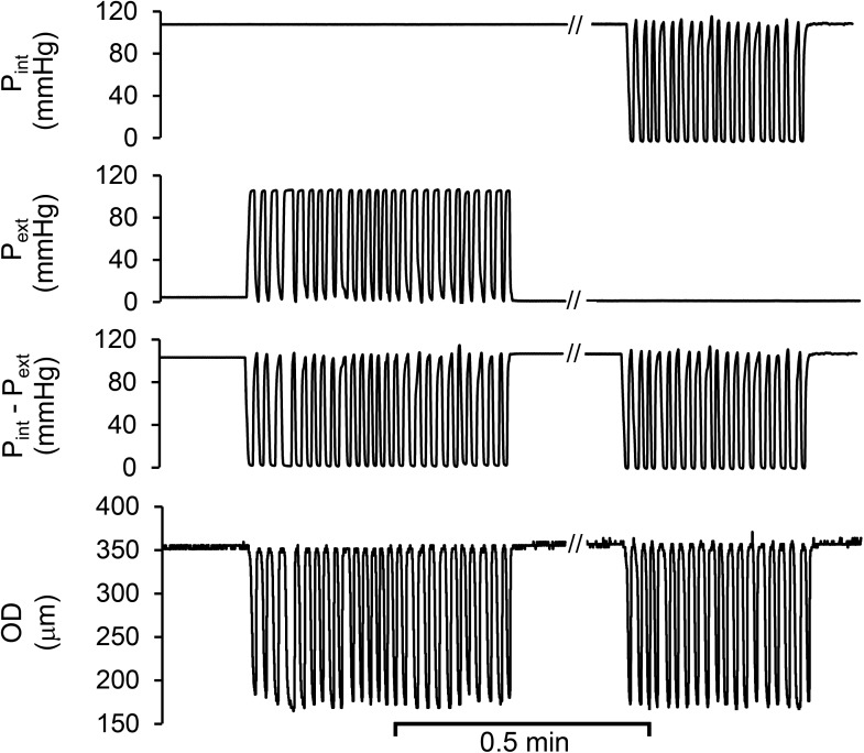
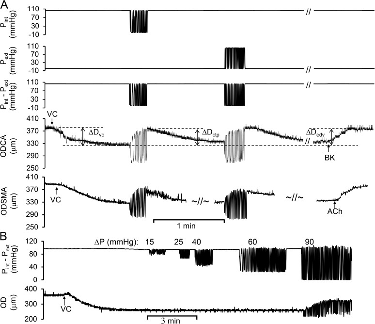
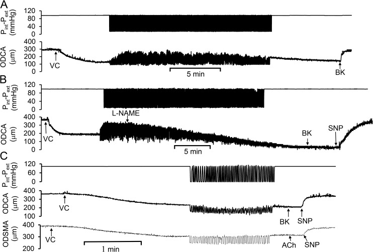
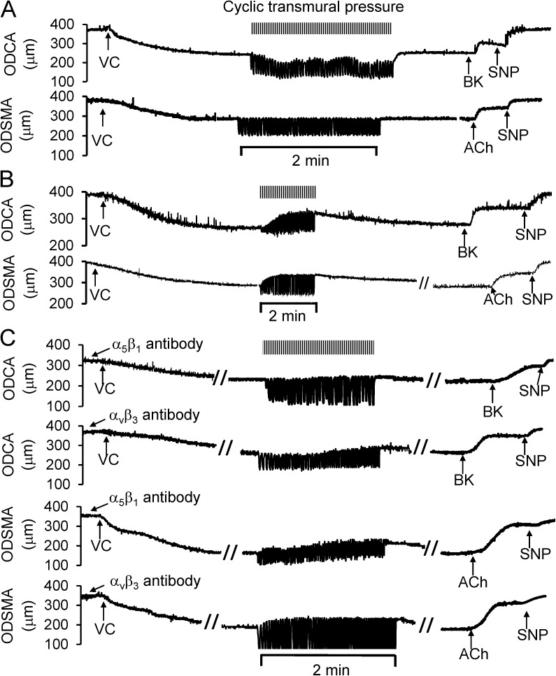
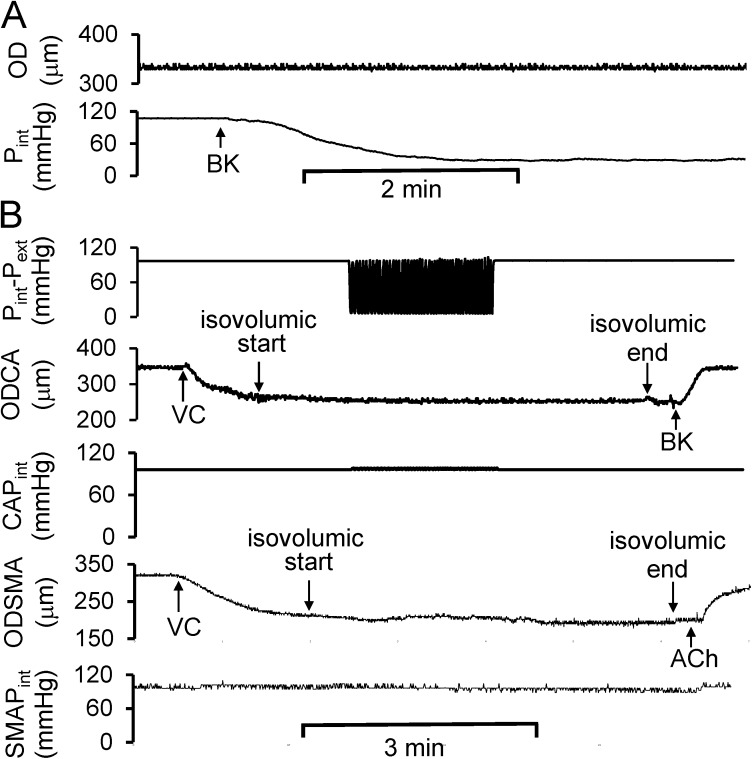
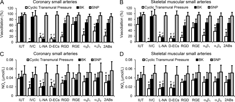
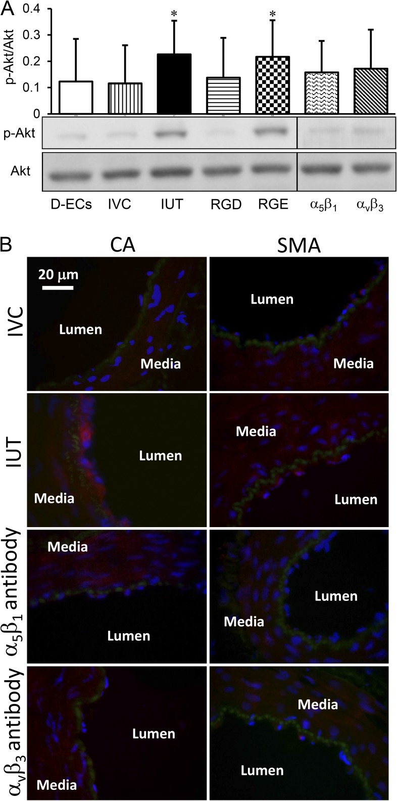
Cardiac and skeletal muscle contraction lead to compression of intramuscular arterioles, which, in turn, leads to their vasodilation (a process that may enhance blood flow during muscle activity). Although endothelium-derived nitric oxide (NO) has been implicated in compression-induced vasodilation, the mechanism whereby arterial compression elicits NO production is unclear. We cannulated isolated swine (n = 39) myocardial (n = 69) and skeletal muscle (n = 60) arteriole segments and exposed them to cyclic transmural pressure generated by either intraluminal or extraluminal pressure pulses to simulate compression in contracting muscle. We found that the vasodilation elicited by internal or external pressure pulses was equivalent; moreover, vasodilation in response to pressure depended on changes in arteriole diameter. Agonist-induced endothelium-dependent and -independent vasodilation was used to verify endothelial and vascular smooth muscle cell viability. Vasodilation in response to cyclic changes in transmural pressure was smaller than that elicited by pharmacological activation of the NO signaling pathway. It was attenuated by inhibition of NO synthase and by mechanical removal of the endothelium. Stemming from previous observations that endothelial integrin is implicated in vasodilation in response to shear stress, we found that function-blocking integrin α5β1 or αvβ3 antibodies attenuated cyclic compression-induced vasodilation and NOx (NO(-)2 and NO(-)3) production, as did an RGD peptide that competitively inhibits ligand binding to some integrins. We therefore conclude that integrin plays a role in cyclic compression-induced endothelial NO production and thereby in the vasodilation of small arteries during cyclic transmural pressure loading.


Figures

Figure 1.
Figure 1.
Typical diameter variation of coronary arterial segment without pre-constriction was caused by cyclic transmural pressures that were generated by either intraluminal or extraluminal pressure variation. Pint, the intraluminal pressure constant at ∼90 mmHg and later cyclically varied from ∼90 to 0 mmHg; Pext, extraluminal pressure cyclically varied from 0 to ∼90 mmHg and later maintained constant at 0 mmHg; Pint–Pext, the cyclic transmural pressure calculated by the difference of Pint and Pext. OD, outer diameter of the segment was automatically tracked during cyclic transmural pressure. The defined abbreviations here are applied in all subsequent figures.
Figure 2.
Figure 2.
The cyclic transmural pressure induced the vasodilation in the precontracted coronary and skeletal muscular small arterial segments. Pint was cyclically varied from ∼90 to 0 mmHg and later maintained constant at ∼90 mmHg. Pext was maintained constant at 0 mmHg and later cyclically varied from 0 to ∼90 mmHg. Pint–Pext was similar regardless of intraluminal or extraluminal variations. (A) The vasodilation in coronary and skeletal muscle arteries. ODCA was the OD of coronary arterial segment, which was tracked in real time during variations of transmural pressure. VC, vasoconstrictor (endothelin-1); ΔDvc, diameter change by vasoconstrictor; ΔDctp, peak diameter change by cyclic transluminal pressure; ΔDedv, peak diameter change by endothelium-dependent vasodilator; BK and ACh, bradykinin and acetylcholine (endothelium-dependent vasodilator). ODSMA was the OD of skeletal muscular arterial segment, which was tracked in real time during variations of transmural pressure. Vasodilation (%) = ΔDctp/ΔDvc. The defined abbreviations here are applied in all subsequent figures. (B) The vasodilation in response to the magnitude of the cyclic transmural pressure.
Figure 3.
Figure 3.
Endothelial role in the cyclic transmural pressure–induced vasodilation. The cyclic transmural pressure induced the vasodilation in precontracted coronary and skeletal muscular small arterial segments, and the vasodilation was inhibited by nonselective NOS inhibitor or denuded endothelium. (A) Cyclic transmural pressure–induced vasodilation persisted for at least 20 min. (B) Administration of L-NAME caused a dynamic vasoconstriction during cyclic transmural pressure. SNP, sodium nitroprusside (a NO donor). (C) Cyclic transmural pressure–induced vasodilation was inhibited by the mechanical removal of endothelium by a vessel diameter–matched wire.
Figure 4.
Figure 4.
Role of integrins in the cyclic transmural pressure–induced vasodilation. (A) RGD peptide (GRGDSP) incubated with the vessel segments for 60 min prevented the vasodilation induced by the cyclic transmural pressure. (B) Control RGE peptide (GRGESP) did not affect the cyclic transmural pressure–induced vasodilation. (C) Function-blocking integrin α5β1 or αvβ3 antibodies compromised the cyclic transmural pressure–induced vasodilation. The top two panels represent the coronary arteries, and bottom two panels represent the skeletal muscle arteries. The vessel segments were incubated with function-blocking integrin α5β1 and αvβ3 antibodies, which were continuously administrated into lumen with a microsyringe pump.
Figure 5.
Figure 5.
Role of circumferential stretch in the cyclic transmural pressure–induced vasodilation. (A) The representation of the decrease in Pint during BK-induced endothelium-dependent vasodilation. Pext was equal to zero. (B) At isovolumic condition, e.g., approximately constant diameter during cyclic transmural pressure, the cyclic transmural pressure failed to elicit integrin-mediated endothelium-dependent vasodilation in both coronary and skeletal muscular small arterial segments. CAPint, intraluminal pressure of coronary small arterial segment; SMAPint, intraluminal pressure of skeletal muscular small arterial segment.
Figure 6.
Figure 6.
Vasodilation and NOx (nitrate/nitrite) production of coronary and skeletal muscular small arteries were induced by the mechanical or pharmacological stimulations. (A) Vasodilation of coronary small arteries was induced by the cyclic transmural pressure or stimulated by BK, ACh, or SNP after the treatments. (B) Vasodilation of skeletal muscular small arteries was induced by the cyclic transmural pressure or stimulated by BK, ACh, or SNP after the treatments. (C) NOx production of coronary small arteries was exposed to stimulations after treatments. (D) NOx production of skeletal muscular small arteries was exposed to stimulations after treatments. The treatments included intact and untreated vessel (IUT), isovolumic condition (IVC) to maintain the diameter constant during cyclic transmural pressure, L-NAME incubation (L-NA), endothelial cells mechanically removed using a wire (D-ECs), GRGDSP peptide incubation (RGD), GRGESP peptide incubation (RGE), incubation with function-blocking integrin α5β1 antibody (α5β1), incubation with function-blocking integrin αvβ3 antibody (αvβ3), and incubation with function-blocking integrin α5β1 and αvβ3 antibodies (2ABs). The number of the vessel segments in the various groups was as follows: coronary segments: n = 15 for IUT, n = 6 for L-NA, n = 5 for D-ECs, n = 6 for RGD, n = 6 for RGE, n = 5 for IVC, n = 6 for α5β1, n = 5 for αvβ3, and n = 11 for 2ABs; skeletal muscular vessel segments: n = 10 for IUT, n = 5 for L-NA, n = 5 for D-ECs, n = 6 for RGD, n = 5 for RGE, n = 4 for IVC, n = 6 for α5β1, n = 6 for αvβ3, and n = 10 for 2ABs. *, P < 0.05 versus incubations; #, P < 0.05 versus stimulations. Error bars represent mean ± SD.
Figure 7.
Figure 7.
Akt phosphorylation of the small arterial segments stimulated by the cyclic transmural pressure after treatments. (A) Expression of Akt phosphorylation. Akt, protein kinase B; p-Akt, phosphorylated Akt. The treatments included the endothelial cells mechanically denuded using a wire (D-ECs), intact and untreated vessel segment (IUT), isovolumic condition (IVC) to maintain diameter constant during the cyclic transmural pressure, GRGDSP peptide incubation (RGD), GRGESP peptide incubation (RGE), incubation with function-blocking integrin α5β1 antibody (α5β1), and incubation with function-blocking integrin αvβ3 antibody (αvβ3). *, P < 0.05 versus IVC. Error bars represent mean ± SEM. (B) Immunofluorescence to verify Akt phosphorylation in endothelial cells. Fluorescent secondary antibody (Alexa Fluor 546; red) indicated that the signal of Akt phosphorylation was stronger in endothelial cells than that in the medial layer of both coronary small arteries (CA) and skeletal muscular small arteries (SMA) in IUT, and no difference between endothelial cells and the medial layer in both CA and SMA at IVC, α5β1 antibody inhibition, or αvβ3 antibody inhibition. Internal elastic lamina was indicated by green and the nucleus by blue.

Similar articles

Cited by

References

    1. Awolesi M.A., Widmann M.D., Sessa W.C., and Sumpio B.E.. 1994. Cyclic strain increases endothelial nitric oxide synthase activity. Surgery. 116:439–444, discussion:444–445. - PubMed
    1. Awolesi M.A., Sessa W.C., and Sumpio B.E.. 1995. Cyclic strain upregulates nitric oxide synthase in cultured bovine aortic endothelial cells. J. Clin. Invest. 96:1449–1454. 10.1172/JCI118181 - DOI - PMC - PubMed
    1. Buckwalter J.B., Ruble S.B., Mueller P.J., and Clifford P.S.. 1998. Skeletal muscle vasodilation at the onset of exercise. J. Appl. Physiol. 85:1649–1654. - PubMed
    1. Chiu J.J., and Chien S.. 2011. Effects of disturbed flow on vascular endothelium: pathophysiological basis and clinical perspectives. Physiol. Rev. 91:327–387. 10.1152/physrev.00047.2009 - DOI - PMC - PubMed
    1. Clifford P.S., Kluess H.A., Hamann J.J., Buckwalter J.B., and Jasperse J.L.. 2006. Mechanical compression elicits vasodilatation in rat skeletal muscle feed arteries. J. Physiol. 572:561–567. 10.1113/jphysiol.2005.099507 - DOI - PMC - PubMed

Publication types

LinkOut - more resources