. 2015 Oct 9;290(41):24835-43.
doi: 10.1074/jbc.M115.650101. Epub 2015 Aug 31.

Pertussis Toxin Exploits Specific Host Cell Signaling Pathways for Promoting Invasion and Translocation of Escherichia coli K1 RS218 in Human Brain-derived Microvascular Endothelial Cells

Affiliations

Pertussis Toxin Exploits Specific Host Cell Signaling Pathways for Promoting Invasion and Translocation of Escherichia coli K1 RS218 in Human Brain-derived Microvascular Endothelial Cells

Sascha Karassek et al. J Biol Chem. .

Abstract

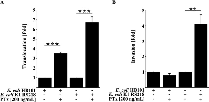
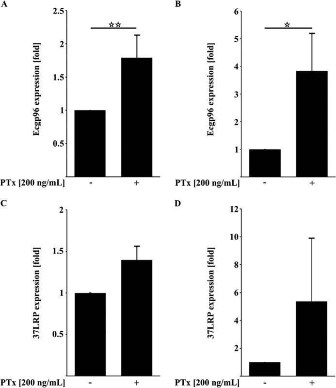
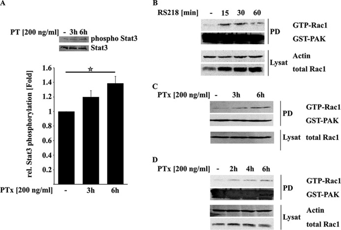
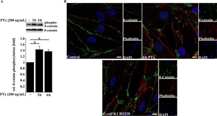
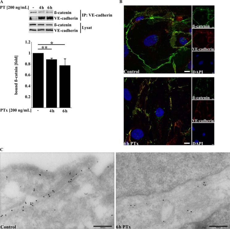
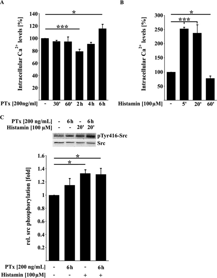
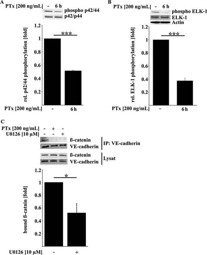
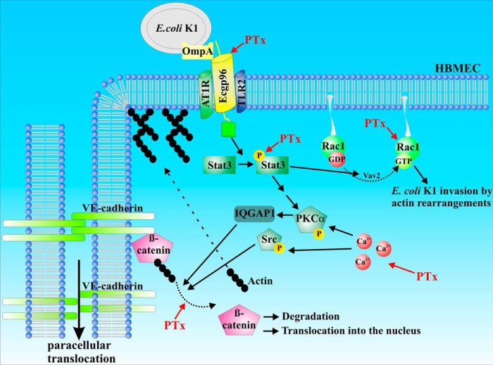
Pertussis toxin (PTx), an AB5 toxin and major virulence factor of the whooping cough-causing pathogen Bordetella pertussis, has been shown to affect the blood-brain barrier. Dysfunction of the blood-brain barrier may facilitate penetration of bacterial pathogens into the brain, such as Escherichia coli K1 (RS218). In this study, we investigated the influence of PTx on blood-brain barrier permissiveness to E. coli infection using human brain-derived endothelial HBMEC and TY10 cells as in vitro models. Our results indicate that PTx acts at several key points of host cell intracellular signaling pathways, which are also affected by E. coli K1 RS218 infection. Application of PTx increased the expression of the pathogen binding receptor gp96. Further, we found an activation of STAT3 and of the small GTPase Rac1, which have been described as being essential for bacterial invasion involving host cell actin cytoskeleton rearrangements at the bacterial entry site. In addition, we showed that PTx induces a remarkable relocation of VE-cadherin and β-catenin from intercellular junctions. The observed changes in host cell signaling molecules were accompanied by differences in intracellular calcium levels, which might act as a second messenger system for PTx. In summary, PTx not only facilitates invasion of E. coli K1 RS218 by activating essential signaling cascades; it also affects intercellular barriers to increase paracellular translocation.

Keywords: Bordetella pertussis; Escherichia coli (E. coli); VE-cadherin; beta-catenin (B-catenin); endothelial dysfunction; pertussis toxin; translocation.


Figures

FIGURE 1.
FIGURE 1.
PTx increases invasion and translocation of E. coli K1 RS218. A, pretreatment of HBMEC with PTx (200 ng/ml) for 6 h significantly increases the translocation rate of E. coli HB101 by 3.52 ± 0.13-fold and E. coli K1 RS218 by 6.72 ± 0.55-fold, respectively. B, 6-h PTx pretreatment (200 ng/ml) of HBMEC significantly increases the invasion rate of the pathogenic E. coli K1 RS218 strain by 4.11 ± 0.59-fold compared with untreated HBMEC. No significant changes were observed for the nonpathogenic E. coli HB101 strain. Bars, mean ± S.E. (error bars) of three independent experiments performed in triplicate. **, p < 0.01; ***, p < 0.001 (determined by ANOVA followed by Bonferroni post hoc test).
FIGURE 2.
FIGURE 2.
PTx up-regulates the expression of the E. coli-binding receptors gp96 but not 37LRP. A and B, quantitative RT-PCR analysis of PTx-treated HBMEC and TY10 cells (200 ng/ml, 6 h) showed significantly increased gp96 mRNA levels. C and D, application of PTx to HBMEC and TY10 cells (200 ng/ml, 6 h) led to no significant changes in 37LRP mRNA levels. Bars, mean ± S.E. of at least six independent experiments performed in duplicate. **, p < 0.01; ***, p < 0.001 (determined by ANOVA).
FIGURE 3.
FIGURE 3.
PTx increases STAT3 and Rac1 activity. A, treatment of serum-deprived HBMEC with PTx (200 ng/ml, 3 or 6 h) led to an increased phosphorylation of Stat3 Tyr-705. B–D, infection of HBMEC with E. coli K1 RS218 activates Rac1 within the first 30 min (B). In contrast, PTx activates Rac1 in HBMEC (C) and TY10 cells (D) significantly after 6 h of stimulation. Bars, mean ± S.E. (error bars) of at least six independent experiments performed in duplicate. Western blots of Rac1 pull-downs give representative results of three independent experiments. **, p < 0.01; ***, p < 0.001 (determined by ANOVA).
FIGURE 4.
FIGURE 4.
PTx increases β-catenin phosphorylation and alters its membrane localization. A, application of PTx (200 ng/ml for 3 or 6 h) significantly increased β-catenin phosphorylation at Thr-41/Ser-45. B, confocal image of TY10 cells stained for β-catenin (Alexa Fluor 488), phalloidin (Alexa Fluor 594), and DAPI either without treatment or with PTx treatment (200 ng/ml, 6 h) or infected with E. coli K1 RS218 (multiplicity of infection of 100, 90 min) shows that β-catenin localization at the cellular membrane is disturbed in the case of PTx application (top right) and E. coli K1 RS218 infection (bottom). In addition, gaplike structures appear in PTx-stimulated and E. coli K1 RS218-infected samples. Scale bar, 10 μm. Bars, mean ± S.E. (error bars) of at least three independent experiments performed in duplicate. *, p < 0.05 (determined by ANOVA).
FIGURE 5.
FIGURE 5.
PTx decreases β-catenin/VE-cadherin interaction and disturbs VE-cadherin membrane localization. A, PTx decreases the amount of β-catenin bound to precipitated VE-cadherin in TY10 after 4–6 h significantly. B, immunofluorescence double staining of β-catenin (Alexa Fluor 594) and VE-cadherin (Alexa Fluor 488) followed by confocal imaging shows a strong colocalization of both proteins at the cellular membrane, which is disrupted after application of PTx (200 ng/ml, 6 h). Scale bar, 10 μm. C, double immunogold labeling of ultra-cryo-sections of TY10 cells shows in control cells a strong localization of VE-cadherin (15-nm gold) and β-catenin (10-nm gold) at outer cellular membranes (left). Application of PTx (200 ng/ml, 6 h) decreases the amount of detectable VE-cadherin and β-catenin at the outer cellular membranes. Bars, mean ± S.E. of at least three independent experiments performed in duplicate. *, p < 0.05; **, p < 0.01 (determined by ANOVA).
FIGURE 6.
FIGURE 6.
PTx influences Ca2+ levels but is not able to activate the protein kinase Src in TY10 cells. A, upon application of PTx (200 ng/ml), Ca2+ levels are significantly decreased after 2 h before they rise again to be significantly increased after 6 h of toxin application. B, compared with the PTx-induced changes in intracellular Ca2+ levels, histamine induces a fast and strong increase of Ca2+ in TY10 cells (5 min, 100 μm). C, Western blotting studies showed that PTx (200 ng/ml, 6 h) has no influence on the activation phosphorylation of the protein kinase Src in contrast to histamine (100 μm, 20 min). Bars, mean ± S.E. (error bars) of at least three independent experiments performed in duplicate. *, p < 0.05; ***, p < 0.001 (determined by ANOVA)
FIGURE 7.
FIGURE 7.
PTx decreases p42/44 MAPK activity, which is sufficient to reduce β-catenin/VE-cadherin interaction. A and B, PTx (200 ng/ml, 6 h) significantly decreased p42/44 MAPK activation phosphorylation at Thr-202/Tyr-204 (A) as well as activation phosphorylation of the p42/44 MAPK downstream target ELK-1 at Ser-383 (B). C, inhibition of p42/44 MAPK by U0126 (10 μm, 6 h) reduced the amount of β-catenin bound to VE-cadherin. Bars, mean ± S.E. (error bars) of at least three independent experiments performed in duplicate. *, p < 0.05; ***, p < 0.001 (determined by ANOVA).
FIGURE 8.
FIGURE 8.
Schematic representation of PTx-affected pathways in transcellular versus paracellular translocation. Based on the data obtained in this study, we hypothesize that PTx preactivates essential signaling pathways for E. coli K1 RS218 invasion and translocation. Up-regulation of gp96 and activation of STAT3 and Rac1, respectively, increase invasive processes. Dissociation of β-catenin and VE-cadherin weakens adherens junctions and enhances paracellular translocation.

Similar articles

Cited by

References

    1. Geier D. A., Geier M. R. (2004) An evaluation of serious neurological disorders following immunization: a comparison of whole-cell pertussis and acellular pertussis vaccines. Brain Dev. 26, 296–300 - PubMed
    1. Heininger U., Kleemann W. J., Cherry J. D., and Sudden Infant Death Syndrome Study Group (2004) A controlled study of the relationship between Bordetella pertussis infections and sudden unexpected deaths among German infants. Pediatrics 114, e9–e15 - PubMed
    1. Grant C. C., McKay E. J., Simpson A., Buckley D. (1998) Pertussis encephalopathy with high cerebrospinal fluid antibody titers to pertussis toxin and filamentous hemagglutinin. Pediatrics 102, 986–990 - PubMed
    1. Seidel G., Böcker K., Schulte J., Wewer C., Greune L., Humberg V., Schmidt M. A. (2011) Pertussis toxin permeabilization enhances the traversal of Escherichia coli K1, macrophages, and monocytes in a cerebral endothelial barrier model in vitro. Int. J. Med. Microbiol. 301, 204–212 - PubMed
    1. Brückener K. E., el Bayâ A., Galla H. J., Schmidt M. A. (2003) Permeabilization in a cerebral endothelial barrier model by pertussis toxin involves the PKC effector pathway and is abolished by elevated levels of cAMP. J. Cell Sci. 116, 1837–1846 - PubMed

Publication types

MeSH terms