. 2015 Oct 16;290(42):25579-94.
doi: 10.1074/jbc.M115.648204. Epub 2015 Aug 31.

Pore-forming Activity of the Escherichia coli Type III Secretion System Protein EspD

Affiliations

Pore-forming Activity of the Escherichia coli Type III Secretion System Protein EspD

Abhishek Chatterjee et al. J Biol Chem. .

Abstract

Enterohemorrhagic Escherichia coli is a causative agent of gastrointestinal and diarrheal diseases. Pathogenesis associated with enterohemorrhagic E. coli involves direct delivery of virulence factors from the bacteria into epithelial cell cytosol via a syringe-like organelle known as the type III secretion system. The type III secretion system protein EspD is a critical factor required for formation of a translocation pore on the host cell membrane. Here, we show that recombinant EspD spontaneously integrates into large unilamellar vesicle (LUV) lipid bilayers; however, pore formation required incorporation of anionic phospholipids such as phosphatidylserine and an acidic pH. Leakage assays performed with fluorescent dextrans confirmed that EspD formed a structure with an inner diameter of ∼2.5 nm. Protease mapping indicated that the two transmembrane helical hairpin of EspD penetrated the lipid layer positioning the N- and C-terminal domains on the extralumenal surface of LUVs. Finally, a combination of glutaraldehyde cross-linking and rate zonal centrifugation suggested that EspD in LUV membranes forms an ∼280-320-kDa oligomeric structure consisting of ∼6-7 subunits.

Keywords: EHEC; EPEC; Escherichia coli (E. coli); EspD; bacterial pathogenesis; membrane protein; protein translocation; type III secretion system (T3SS).


Figures

FIGURE 1.
FIGURE 1.
Schematic architecture of EHEC EspD. Bioinformatic structural analysis of EspD predicted two amphipathic regions (Amph I and Amph II, residues 1–83), two coiled-coil motifs (COIL I residues 138–171 and COIL II residues 334–379), and two transmembrane helices (residues 176–200 and 227–251). Cylinders represent predicted helical structures, and thick lines correspond to β-strand or random coil structures. Gray squares located in the loop spanning residues 200–227 represent aspartate, glutamate, and lysine residues, and tryptophan residues are denoted by a W.
FIGURE 2.
FIGURE 2.
Purification of recombinant EspD. Recombinant EspD was purified as an EspD-chitin binding domain fusion protein. Mid-log phase cultures (uninduced) were treated with isopropyl β-d-thiogalactopyranoside (induced). EspD bound to the chitin column was cleaved with DTT and recovered in the eluate (chitin eluate). EspD was further purified with Triton X-114 detergent phase separation (Triton X-114). Aliquots were resolved by SDS-PAGE, and proteins were electrotransferred to PVDF membrane and developed by Coomassie Blue dye staining (A) or Western blot analysis using anti-EspD-specific antisera (B).
FIGURE 3.
FIGURE 3.
Phospholipid specificity for EspD binding. EspD was mixed with SUVs composed of single phospholipids (A) or a mixture of phospholipids plus cholesterol in PBS (B) and the SUV bound and free EspD separated by sucrose density flotation centrifugation. C, EspD was incubated with SM/DOPC/DOPS/Chl LUVs at pH 4.5 or pH 7.2, and protein binding was assessed by Western blot analysis following sucrose density centrifugation using anti-EspD antibodies.
FIGURE 4.
FIGURE 4.
Interaction of EspD with lipid bilayers. A, SM/DOPC/DOPS/Chl LUV loaded with EspD at pH 4.5 or 7.2 and purified by sucrose density flotation centrifugation were sequentially extracted with 500 mm NaCl, 100 mm alkaline carbonate, pH 11.5, and 8.0 m urea in PBS and separated into a supernatant (S) or pellet (P) fraction. The interaction with HeLa cell membranes was assessed by incubating cells with recombinant EspD and then subjecting a HeLa crude membrane pellet to the sequential alkaline and urea extraction described above. B, LUVs loaded with EspD at pH 4.5 or 7.2 were extracted at 0 °C with Triton X-100, and the soluble and insoluble fractions were separated by centrifugation as described above. EspD distribution was assessed by Western blot analysis using anti-EspD antisera.
FIGURE 5.
FIGURE 5.
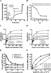
Dye leakage activity of EspD. A, pore formation was evaluated by addition of EspD to SRB-loaded SM/DOPC/DOPS/Chl LUVs at pH 7.2, and dye release was monitored prior to shifting the pH to 4.5. B, effect of pH and phospholipid composition on pore formation was examined by adding EspD (EspD ratio 1:1250) to SRB-loaded SM/DOPC/DOPS/Chl or SM/DOPC/DOPE:Chl LUVs at various pH values and monitoring dye release. C, dependence of DOPS on EspD pore formation at pH 4.5 was assessed using SM/DOPC/DOPS/Chl (45:Y:X:20) membranes with increasing DOPS levels. The dependence of cholesterol on pore formation at pH 4.5 was evaluated using SM/DOPC/DOPS/Chl: (45:Y:20:X) where the cholesterol mol % was varied. D, interaction affinity of EspD with lipid bilayers of various composition was determined by incubating a fixed amount of SUV with increasing concentrations of EspD and separating the free and SUV-associated EspD by sucrose density flotation and quantifying the protein in the bound fraction by ELISA. E, to assess the importance of electrostatic interactions on pore formation, leakage assays were performed in the presence of increasing NaCl concentrations using a constant EspD/phospholipid mole ratio of 1:2500. F, effect of fatty acid composition on pore formation was examined by dye leakage assays at pH 4.5 and pH 7.2 using LUVs composed of SM/DOPC/DOPS/Chl (44:24:12:20) (DO PL) or SM:POPC:POPS:Chl (44:24:12:20) (PO PL) and increasing EspD to phospholipid mole ratios.
FIGURE 6.
FIGURE 6.
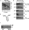
Orientation of EspD in the lipid bilayer. A, capacity of the EspD (WT) and espD Trp-47 (W47), espD Trp-178 (W178), and espD Trp-195 (W195) mutants to bind SM/DOPC/DOPS/Chl LUVs was assessed by Western blot analysis of sucrose density centrifugation fractions. B, pore-forming activity of the espD mutants was assessed by monitoring SRB release from SM/DOPC/DOPS/Chl LUVs at pH 4.5. Insertion of espD Trp-47 (C), espD W Trp-178 (D), or espD Trp-195 (E) into SM/DOPC/DOPS/Chl SUVs membranes at pH 4.5 or 7.2 was examined by intrinsic fluorescence spectroscopy at an excitation wavelength of 295 nm. Spectra were corrected by subtracting traces with SUVs alone. F, depth of tryptophan insertion into the lipid bilayer at pH 4.5 or 7.2 was assessed by acrylamide-induced fluorescence quenching. For fluorescence spectroscopy experiments, an EspD/PL ratio of 1:500 was used.
FIGURE 7.
FIGURE 7.
Membrane topology of EspD. A, EspD digested with clostripain for various times was tested for dye leakage activity (values below panel) at pH 4.5 (EspD/PL, 1:250). B, limited clostripain EspD digest product is illustrated. C, membrane binding activity of the EspD(95–295) fragment was assessed by sucrose density centrifugation. D, interaction of EspD with LUV membranes at pH 4.5 and 7.2 was evaluated by tryptic digest in the absence or presence of Triton X-100. Degradation of EspD was monitored using anti-EspD antisera.
FIGURE 8.
FIGURE 8.
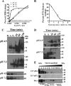
Characterization of the EspD pore. A, effect of pH and EspD concentration on pore formation was examined by titrating SRB SM/DOPC/DOPS/Chl LUVs with increasing concentrations of EspD at pH 4.0, 5.5, and 7.2, and the percent dye release was plotted as a function of the EspD/phospholipid ratio. B, diameter of the EspD pore formed at pH 4.5 was determined with LUVs loaded with carboxyfluorescein or FITC labeled 4–70-kDa dextrans. The oligomeric state of EspD on LUVs at pH 4.5 or 7.2 was assessed by glutaraldehyde cross-linking, and complexes were examined by Western blot on a 6% (upper panel) and 10% (lower panel) SDS-polyacrylamide gel (C). D, clostripain fragment of EspD encompassing residues 95–295 were glutaraldehyde cross-linked after loading protein onto LUVs at pH 4.5 or pH 7.2. E, complexes formed by recombinant and native EspD on LUV and HeLa cell membranes were examined by rate zonal centrifugation on a linear sucrose gradient following detergent extraction of complexes.
FIGURE 9.
FIGURE 9.
Model of EspD membrane interaction. A, at pH 7.2 the transmembrane helices and the N-terminal amphipathic region of EspD are postulated to insert into the outer leaflet of the lipid bilayer in a parallel orientation with the coiled-coil motifs (COIL I and COIL II) remaining solvent-exposed and mediate EspD oligomerization through COIL I-COIL I and COIL II-COIL II interactions to form a pre-pore structure. B, conformational changes triggered by an acidic pH are postulated to induce structural rearrangements that involve the penetration of the hairpin loop and formation of a pore resulting in a re-orientation of the transmembrane domains in a configuration that is perpendicular to the lipid bilayer. C, multiple sequence alignments showing the distribution of acidic residues in the hairpin loop region for the EspD homologues from a Salmonella, Pseudomonas, Yersinia, and Shigella.

Similar articles

Cited by

References

    1. Guarner F., Malagelada J. R. (2003) Gut flora in health and disease. Lancet 361, 512–519 - PubMed
    1. Marteau P., Pochart P., Doré J., Béra-Maillet C., Bernalier A., Corthier G. (2001) Comparative study of bacterial groups within the human cecal and fecal microbiota. Appl. Environ. Microbiol. 67, 4939–4942 - PMC - PubMed
    1. Russo T. A., Johnson J. R. (2003) Medical and economic impact of extraintestinal infections due to Escherichia coli: focus on an increasingly important endemic problem. Microbes Infect. 5, 449–456 - PubMed
    1. Kaper J. B., Nataro J. P., Mobley H. L. (2004) Pathogenic Escherichia coli. Nat. Rev. Microbiol. 2, 123–140 - PubMed
    1. Garmendia J., Frankel G., Crepin V. F. (2005) Enteropathogenic and enterohemorrhagic Escherichia coli infections: translocation, translocation, translocation. Infect. Immun. 73, 2573–2585 - PMC - PubMed

Publication types

MeSH terms

LinkOut - more resources