. 2015 Oct 16;290(42):25461-74.
doi: 10.1074/jbc.M115.670984. Epub 2015 Aug 31.

Structure of the Bacillus anthracis Sortase A Enzyme Bound to Its Sorting Signal: A FLEXIBLE AMINO-TERMINAL APPENDAGE MODULATES SUBSTRATE ACCESS

Affiliations

Structure of the Bacillus anthracis Sortase A Enzyme Bound to Its Sorting Signal: A FLEXIBLE AMINO-TERMINAL APPENDAGE MODULATES SUBSTRATE ACCESS

Albert H Chan et al. J Biol Chem. .

Abstract

The endospore forming bacterium Bacillus anthracis causes lethal anthrax disease in humans and animals. The ability of this pathogen to replicate within macrophages is dependent upon the display of bacterial surface proteins attached to the cell wall by the B. anthracis Sortase A ((Ba)SrtA) enzyme. Previously, we discovered that the class A (Ba)SrtA sortase contains a unique N-terminal appendage that wraps around the body of the protein to contact the active site of the enzyme. To gain insight into its function, we determined the NMR structure of (Ba)SrtA bound to a LPXTG sorting signal analog. The structure, combined with dynamics, kinetics, and whole cell protein display data suggest that the N terminus modulates substrate access to the enzyme. We propose that it may increase the efficiency of protein display by reducing the unproductive hydrolytic cleavage of enzyme-protein covalent intermediates that form during the cell wall anchoring reaction. Notably, a key active site loop (β7/β8 loop) undergoes a disordered to ordered transition upon binding the sorting signal, potentially facilitating recognition of lipid II.

Keywords: Bacillus anthracis; SrtA; enzyme mechanism; enzyme regulation; enzyme substrate complex; nuclear magnetic resonance (NMR); protein dynamic; protein structure; sortase A; transpeptidase.


Figures

FIGURE 1.
FIGURE 1.
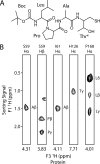
The Boc-LPAT* modifier and NMR data of its complex with BaSrtA. A, the chemical structure of the Boc-LPAT* peptide analog, where T* is (2R,3S)-3-amino-4-mercapto-2-butanol, and Boc is a tert-butyloxycarbonyl protecting group. B, selected panels showing intermolecular NOEs between BaSrtA and the Boc-LPAT* peptide. Panels are taken from a three-dimensional (F1) 13C,15N-filtered, (F2) 13C-edited NOESY-HSQC spectrum of the BaSrtA-LPAT* complex dissolved in deuterated buffer. The identity of the proton from BaSrtA that gives rise to the set of NOEs and its chemical shift are shown at the top and bottom of each panel, respectively. On the right side of each cross-peak the sorting signal peptide proton that is proximal to the protein is indicated.
FIGURE 2.
FIGURE 2.
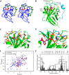
NMR solution structure of the BaSrtA-LPAT* complex. A, stereo image showing the ensemble of 20 lowest energy structures of the BaSrtA-LPAT complex. The protein backbone heavy atoms (blue) and the covalently linked peptide (red) are shown. The N-terminal appendage is colored green. The coordinates were superimposed by aligning the backbone N, Cα, and C atoms of Asp57-Lys210 and Leu, Pro, Ala, and Thr* of the Boc-LPAT* peptide. The backbone and heavy atom coordinates of these residues have a r.m.s. deviation to the average structure of 0.42 and 0.89 Å, respectively. B, ribbon drawing of the energy minimized average structure of the BaSrtA-LPAT* complex. The covalently bound peptide is shown in a red ball-and-stick representation with its amino acids labeled. The N-terminal appendage is colored cyan. C, expanded view of the active site showing how the sorting signal peptide is recognized. The side chains (orange) that participate in the interaction are labeled and shown as sticks. D, expanded view of the active site showing how the N-terminal appendage (cyan) positions over the sorting signal peptide (red). The side chains (orange) that participate in the interaction are labeled and shown as sticks. E, overlay of the 1H-15N HSQC spectra of apo BaSrtA (red) and the BaSrtA-LPAT* complex (blue). F, histogram plot of the compound chemical shift changes (Δδ) for the backbone amide hydrogen and nitrogen atoms of apo BaSrtA after the addition of Boc-LPAT*. Chemical shift changes are calculated by the equation Δδ = ((ΔδH)2 + (ΔδN/6.49)2)1/2, where ΔδH and ΔδN are, respectively, the amide proton and nitrogen chemical shift difference for a given residue in the presence and absence of Boc-LPAT*. The dashed line represents 1 S.D. above the average Δδ of all amino acids. Amino acids experiencing significant chemical shift changes are labeled, and a schematic of secondary structures of the enzyme is shown above the plot.
FIGURE 3.
FIGURE 3.
Mobility of BaSrtA-LPAT* as defined by NMR relaxation data. Panels A–D are scatter plots of the relaxation data for the complex: R1 (A), R2 (B), 1H-15N heteronuclear NOE (C), and general order parameter S2 of the backbone 15N atoms (D) as a function of residue number. The value of S2 ranges from 0 to 1, with a value of 1 indicating that the amide bond is completely immobilized. The secondary structures present in the protein are displayed on top of each plot. E, ribbon drawing of BaSrtA-LPAT* showing the location of residues in which the backbone amide resonances could not be assigned (black spheres), or residues that have Rex values greater than 10 Hz (gray spheres).
FIGURE 4.
FIGURE 4.
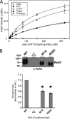
Effects of mutating the N-terminal appendage on enzyme kinetics and protein display in B. anthracis. A, representative curves showing the in vitro hydrolysis kinetics of S59G (open circles), I61A (filled circles), and C187A (open squares) mutants of BaSrtA, as well as the hydrolysis kinetics of wild-type BaSrtA (filled squares). Also shown is the progress curve for mutant of BaSrtA that removes a portion of the N-terminal appendage (BaSrtAΔ64, identical to BaSrtA except residues Asp57-Pro64 are absent). B, the effects of mutations in the N-terminal appendage of sortase on BasC protein display in B. anthracis. Sterne 34F2 strains lacking srtA (▵) or complemented with the indicated srtA and basCFLAG/MH6 on pAHG322 were grown to an A600 of 1, and the harvested cells digested with lysozyme to liberate BasCFLAG/MH6. Cleared lysates were purified via affinity chromatography and the resulting eluates were analyzed for the presence of BasCFLAG/MH6 by immunoblotting with anti-FLAG polyclonal antibodies. A representative blot is shown at the top. Biological replicates were quantified and normalized to the BasCFLAG/MH6 anchored by WT SrtA. Means are plotted with standard error at the bottom panel. * = p < 0.01, n = 3.
FIGURE 5.
FIGURE 5.
Comparison of the structure of BaSrtA in its apo and LPAT* bound states. A, ribbon drawing showing the superposition of the structures of apo BaSrtA (PDB code 2KW8; red) and the BaSrtA-LPAT* complex (green). Key residues are represented as sticks and colored orange (BaSrtA-LPAT*) or magenta (apo BaSrtA). An expanded view of the β7/β8 loop is shown. The loop transitions from a disordered state (represented by dotted line) to an ordered state after binding the substrate. Cys187 is displaced by ∼7 Å away from the substrate binding pocket, facilitating new contacts between the side chains of Val190, Met128, and Ser129. The N-terminal appendage and Boc-LPAT* are removed for clarity. B, as in panel A, but an expanded view of the N-terminal appendage and its interactions with the active site is shown. Ile61 is displaced when the substrate binds such that it only partially buries His126. The backbone atoms of Ile61-Thr186 and Arg196-Lys210 of the two structures align to a r.m.s. deviation of 0.73 Å. C, scatter plot showing the change in the general order parameter S2 after the addition of Boc-LPAT*. A positive number indicates the backbone amide becomes more rigid, whereas a negative number indicates it becomes more disordered. A schematic of secondary structures in the enzyme is shown above the plot. D, histogram plot of the difference in RCI between Boc-LPAT* bound BaSrtA and apo BaSrtA. The dashed line represents 1 S.D. above the average difference in RCI (random coil index) for all amino acids. A positive value indicates that protein likely becomes less structured upon binding the substrate. A schematic of secondary structures of the enzyme is shown above the plot.
FIGURE 6.
FIGURE 6.
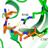
Model of the thioacyl intermediate. Expanded view of the active site in the energy minimized model of the BaSrtA-LPAT thioacyl intermediate. The peptide substrate LPAT (orange) and active site residues Cys187 and Arg196 (green) are shown as sticks. Hydrogen bonds are indicated by yellow dotted lines. The distance between Thr carbonyl oxygen and Arg196 guanidino Cζ is 3.6 Å.
FIGURE 7.
FIGURE 7.
The N-terminal appendage masks a potential binding site for lipid II. The program FTSite was used to predict the binding site for DAP, the amino group containing moiety in the B. anthracis lipid II molecule that is joined to the sorting signal by sortase. The panels show solvent accessible surface representations of BaSrtA-LPAT* (A) and BaSrtA-LPAT* (B) in which residues Asp57-Pro64 of the N-terminal appendage is removed. Only when residues Asp57-Pro64 are removed is a potential binding site located near the active site observed (magenta colored mesh). In the figure, the N-terminal appendage, H2 helix, β4/H3 loop, β7/β8 loop, and active site His126 are colored red, orange, blue, green, and yellow, respectively. The substrate analog LPAT* is shown in stick format. C, as in panel A, but the N-terminal appendage is displayed as a mesh object.

Similar articles

Cited by

References

    1. Mock M., Fouet A. (2001) Anthrax. Annu. Rev. Microbiol. 55, 647–671 - PubMed
    1. Holty J. E., Bravata D. M., Liu H., Olshen R. A., McDonald K. M., Owens D. K. (2006) Systematic review: a century of inhalational anthrax cases from 1900 to 2005. Ann. Intern. Med. 144, 270–280 - PubMed
    1. Zink S. D., Burns D. L. (2005) Importance of srtA and srtB for growth of Bacillus anthracis in macrophages. Infect. Immun. 73, 5222–5228 - PMC - PubMed
    1. Cascioferro S., Totsika M., Schillaci D. (2014) Sortase A: an ideal target for anti-virulence drug development. Microb. Pathog. 77, 105–112 - PubMed
    1. Schneewind O., Missiakas D. (2014) Sec-secretion and sortase-mediated anchoring of proteins in Gram-positive bacteria. Biochim. Biophys. Acta 1843, 1687–1697 - PMC - PubMed

Publication types

MeSH terms