. 2015 Oct 16;290(42):25411-26.
doi: 10.1074/jbc.M115.657775. Epub 2015 Aug 31.

Nuclear Calcium/Calmodulin-dependent Protein Kinase II Signaling Enhances Cardiac Progenitor Cell Survival and Cardiac Lineage Commitment

Affiliations

Nuclear Calcium/Calmodulin-dependent Protein Kinase II Signaling Enhances Cardiac Progenitor Cell Survival and Cardiac Lineage Commitment

Pearl Quijada et al. J Biol Chem. .

Abstract

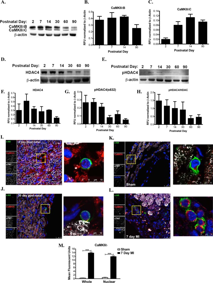
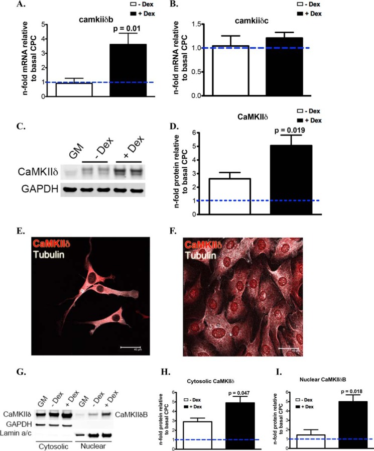
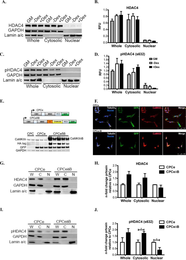
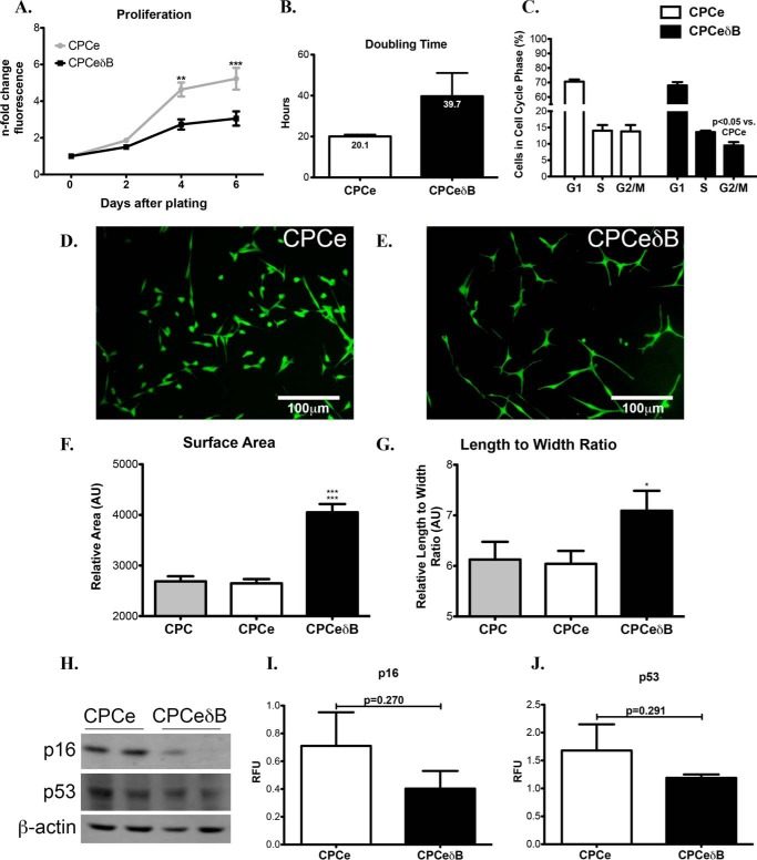
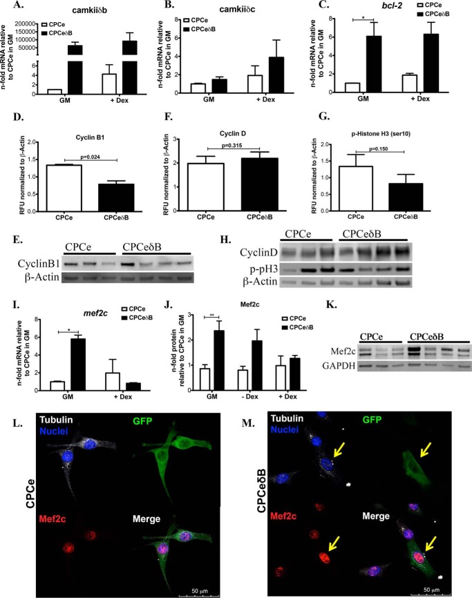
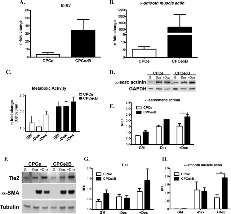
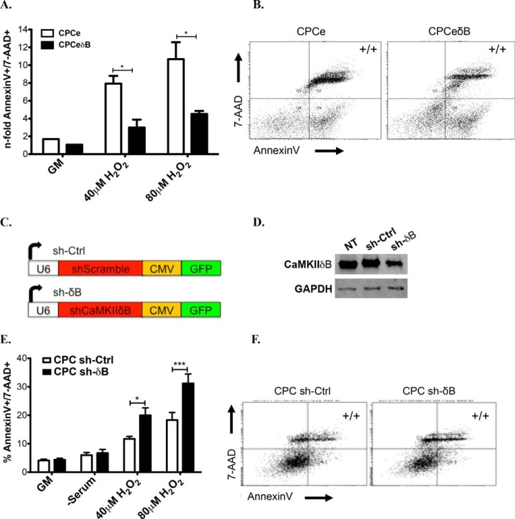
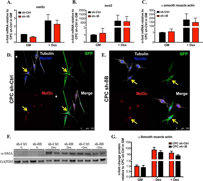
Ca(2+)/Calmodulin-dependent protein kinase II (CaMKII) signaling in the heart regulates cardiomyocyte contractility and growth in response to elevated intracellular Ca(2+). The δB isoform of CaMKII is the predominant nuclear splice variant in the adult heart and regulates cardiomyocyte hypertrophic gene expression by signaling to the histone deacetylase HDAC4. However, the role of CaMKIIδ in cardiac progenitor cells (CPCs) has not been previously explored. During post-natal growth endogenous CPCs display primarily cytosolic CaMKIIδ, which localizes to the nuclear compartment of CPCs after myocardial infarction injury. CPCs undergoing early differentiation in vitro increase levels of CaMKIIδB in the nuclear compartment where the kinase may contribute to the regulation of CPC commitment. CPCs modified with lentiviral-based constructs to overexpress CaMKIIδB (CPCeδB) have reduced proliferative rate compared with CPCs expressing eGFP alone (CPCe). Additionally, stable expression of CaMKIIδB promotes distinct morphological changes such as increased cell surface area and length of cells compared with CPCe. CPCeδB are resistant to oxidative stress induced by hydrogen peroxide (H2O2) relative to CPCe, whereas knockdown of CaMKIIδB resulted in an up-regulation of cell death and cellular senescence markers compared with scrambled treated controls. Dexamethasone (Dex) treatment increased mRNA and protein expression of cardiomyogenic markers cardiac troponin T and α-smooth muscle actin in CPCeδB compared with CPCe, suggesting increased differentiation. Therefore, CaMKIIδB may serve as a novel modulatory protein to enhance CPC survival and commitment into the cardiac and smooth muscle lineages.

Keywords: Ca2+/calmodulin-dependent protein kinase II (CaMKII); cell death; cell differentiation; histone deacetylase 4 (HDAC4); stem cells.


Figures

FIGURE 1.
FIGURE 1.
CaMKIIδ is expressed in CPCs during post-natal growth and is up-regulated after pathological stress. A, CaMKIIδB (top band) and CaMKIIδC (bottom band) protein expression in whole mouse heart lysates at increasing postnatal days. B, quantitation of CaMKIIδB and C, CaMKIIδC expression levels represented as relative fluorescent units (RFU) normalized to β-actin. D, total HDAC4 levels in the post-natal mouse heart. E, phosphorylated HDAC4 on the serine 632 in the post-natal and adult mouse heart. F, quantitation of HDAC4 expression normalized to β-actin represented as RFU. G, quantitation of pHDAC4 expression normalized to β-actin represented as RFU. H, phosphorylated HDAC4 over total HDAC4 in the post-natal and adult mouse heart. I, 2-day-old and J, 30-day-old hearts visualized for expression and localization of CaMKIIδ in myocytes and c-kit+ CPCs. K, CaMKIIδ labeling in CPCs in sham-operated mice or L, following MI surgery for 7 days. M, quantitation of CaMKIIδ expression and localization (whole or nuclear) in endogenous CPCs in sham and 7 day MI-treated mice. Mice are 3 months of age prior to MI surgery. Cardiac troponin T (cTNT) and TO-PRO-3 iodide were used to label myocardium and nuclei, respectively. ***, p < 0.0001 Sham versus 7 Day MI.
FIGURE 2.
FIGURE 2.
CaMKIIδB localization to the nucleus is increased in CPCs during commitment. A, CaMKIIδB and B, CaMKIIδC mRNA in CPCs with and without dexamethasone (Dex) stimulation for 7 days represented as a fold change relative to CPCs maintained in growth medium (GM). C, CaMKIIδ protein levels in whole cell lysates with and without Dex stimulation. D, quantitation of total CaMKIIδ levels relative to GM-treated CPCs. E, CaMKIIδ protein is primarily cytosolic in CPCs without differentiation stimulus. F, CaMKIIδ increases in expression and localizes to the nuclear compartment of CPCs after 6 days of Dex treatment. G, CaMKIIδ expression in the cytosolic and nuclear fractions of CPCs after induction with differentiation medium. H, quantitation of cytosolic CaMKIIδ and I, nuclear CaMKIIδB. Graphs are represented as a fold change relative to basal CPCs in cytosolic and nuclear fractions. GAPDH is probed as a loading control for immunoblotting. p values compare CPCs in 10% FBS α-MEM without Dex to CPCs treated with Dex.
FIGURE 3.
FIGURE 3.
CaMKIIδB localizes to the nucleus and inhibits HDAC4 in CPCs undergoing cardiogenic commitment. A, immunoblot of total HDAC4 and protein levels in whole, cytosolic and nuclear fractions of CPCs treated with GM, −Dex, or + Dex supplemented medium. B, quantitation of total HDAC4. C, immunoblot of pHDAC4 (s632) protein levels in whole, cytosolic, and nuclear fractions of CPCs treated with GM, −Dex, or + Dex-supplemented medium. D, quantitation of pHDAC4 (s632). Immunoblots are presented as a fold change relative to CPCs in GM after normalization to GAPDH or Lamin A (C terminus). E, lentiviral constructs to establish CPCe and CPCeδB lines (top). CPCeδB lines overexpress CaMKIIδB as well as the HA tag. CPCe overexpress eGFP. GAPDH was probed as a loading control (bottom). F, CPCe (top) and CPCeδB (bottom) fluorescent images. Cells were stained with antibodies toward GFP and CAMKIIδ. Cell morphology and nuclei were visualized by staining for tubulin and with DAPI, respectively. G, CPCe and CPCeδB lines probed for total HDAC4 levels by immunoblot and H, quantitation in whole cell lysates, cytosolic and nuclear fractions. I, phosphorylated HDAC4 on the serine 632 protein levels in CPCe or CPCeδB by immunoblot and J, quantitated from whole cell lysates,cytosolic, and nuclear fractions. Immunoblots are probed with GAPDH to normalize for protein loading of the whole and cytosolic lysates. Lamin A (C terminus) antibody was utilized to normalize for loading of the nuclear fraction. Total and phosphorylated HDAC4 are represented as a fold change relative to CPCe. *, p < 0.05 CPCe versus CPCeδB. W, whole cell; C, cytosolic fraction; and N, nuclear fraction.
FIGURE 4.
FIGURE 4.
Nuclear CaMKIIδ overexpression in CPCs promotes increases in cellular size and decreased proliferation. A, CPCeδB has decreased proliferative capacity relative to CPCe based on a fluorescent nucleic acid stain measured at 2, 4, and 6 days after seeding equal cell numbers. ** and *** p < 0.01 and p < 0.0001 CPCeδB relative to CPCe. B, CPCeδB populations shows an increased doubling time relative to CPCe. C, cell cycle analysis using flow cytometry in CPCe and CPCeδB. D, fluorescent images of CPCe and E, CPCeδB populations in growth media. F, CPCeδB shows an increase in relative cell surface area. ***, p < 0.0001 CPCeδB relative to CPC and CPCe. G, CPCeδB shows an increase in length to width ratio relative to non-modified CPCs and CPCe. *, p < 0.01 CPCeδB relative to CPCe. H, expression of senescence markers p16 and p53 by immunoblot. β-Actin was probed as a loading control. I, quantitation of p16 and J, p53 in CPCe and CPCeδB populations.
FIGURE 5.
FIGURE 5.
CaMKIIδB overexpression in CPCs enhances expression of pro-survival and cardiac lineage commitment markers and down-regulates proteins involved in mitotic progression. A, CaMKIIδB; B, CaMKIIδC; C, Bcl-2 gene expression in CPCe and CPCeδB during growth conditions or with the addition of dex. Values are represented as n-fold mRNA relative to CPCe in GM and after normalization to ribosomal 18S. D, cyclin B1 protein quantitation. E, cyclin B1 protein immunoblot representation. F, cyclin D and G, p- histone H3 (Ser-10) quantitation. H, cyclin D and p-Histone H3 immunoblot representation. Values are represented as RFU after normalizing to loading control β-Actin. I. Mef2c gene expression in CPCe and CPCeδB during growth conditions or with the addition of dex. Values are represented as n-fold mRNA relative to CPCe in GM and after normalization to ribosomal 18S. *, p < 0.05. J, Mef2c protein expression in CPCs cultured in GM, medium without the addition of dex or with the addition of dex. Values are represented as n-fold mRNA relative to CPCe in GM and after normalization to loading control GAPDH. **, p < 0.01. K, immunoblot representation of Mef2c in CPCe and CPCeδB maintained in GM conditions alone, quantitation in J. n = 3 CPCe and n = 4 C CPCeδB were analyzed for immunoblotting. L and M, Mef2c protein intensity within nuclei of CPCe and CPCeδB. M, CPCeδB identified by GFP staining express higher Mef2c in the nucleus. Cell morphology and nuclei were visualized by staining for tubulin and with DAPI, respectively.
FIGURE 6.
FIGURE 6.
CaMKIIδB overexpression in CPCs increases cell commitment toward the cardiomyogenic lineage. A, CPCeδB stimulated with Dex for 6 days show increased expression of cardiogenic marker cardiac troponin T (tnnt3) and B. α-smooth muscle actin based on mRNA expression. C, cardiac marker α-sarcomeric expression and D, quantitation in CPCe and CPCeδB in normal growth conditions (G), −Dex and +Dex. E, metabolic activity under GM, −Dex, or +Dex growth conditions. F, endothelial (Tie2) and smooth muscle (α-SMA) were probed in CPCe and CPCeδB grown in normal growth conditions (G), −Dex or +Dex. G, quantitation of Tie2 and H. α-SMA, which is up-regulated in Dex-treated CPCeδB. Values are represented as relative fluorescent units (RFU) normalized to GAPDH or tubulin. *, p < 0.05 and **, p < 0.01 CPCe versus CPCeδB.
FIGURE 7.
FIGURE 7.
CaMKIIδB expression in CPCs antagonizes apoptotic cell death after oxidative stress stimuli. A, quantitation of Annexin V and 7-AAD double positive cells (to label for cells in late apoptosis) in B. CPCe and C, CPCeδB after incubation in growth medium (GM), 40 or 80 μm H2O2. Values are represented as a fold change relative to GM-treated cells. D, schematic of constructs (top) and confirmation of knockdown of CaMKIIδB in CPCs relative to controls (NT: non-treated and sh-Ctrl). E, quantitation of late apoptosis in CPCs transfected with F, small hairpin (sh) control and G, Sh to knockdown CaMKIIδB and subjected to GM, 40, or 80 μm H2O2. Values are represented as a percentage based on flow cytometric analysis.
FIGURE 8.
FIGURE 8.
Knockdown of CaMKIIδB does not impair commitment of CPCs toward the cardiac or smooth muscle phenotype. A, cardiac markers Mef2c; B, cardiac troponin T (tnnt3); and C, smooth muscle marker α-smooth muscle actin gene expression in CPC sh-Ctrl and CPC sh-δB during growth conditions and after the addition of dex. Values are represented as fold change mRNA relative to CPC sh-Ctrl in GM after normalizing to ribosomal 18. D, CPC sh-Ctrl and E, CPC sh-δB fluorescent images after staining for Mef2c protein. Cells were maintained in GM. Cells were stained with antibodies toward GFP to identify cells that were successfully transduced with the sh-based lentivirus. Cell morphology and nuclei were visualized by staining for tubulin and with DAPI, respectively. F, immunoblotting for α-smooth muscle actin in CPC sh-Ctrl and CPC sh-δB in GM, medium without dex, or medium with dex. G, quantitation of α-smooth muscle actin represented as a fold change relative to CPC sh-Ctrl in GM and after normalizing to GAPDH.
FIGURE 9.
FIGURE 9.
Knockdown of CaMKIIδB in CPCs prevents the up-regulation of pro-apoptotic molecules and promotes properties of cellular senescence. A, CaMKIIδB; B, CaMKIIδC; and C, Bcl-2 gene expression in CPC sh-Ctrl and CPC sh-δB during growth conditions or with the addition of dex. Values are represented as n-fold mRNA relative to CPC sh-Ctrl in GM and after normalization to ribosomal 18S. **, p < 0.01. D, proliferation measured using a direct nucleic acid fluorescent dye. Fluorescent values are represented as a fold change relative to day of plating (Day 0). E, cell doubling time in hours. F, cell cycle analysis using propidium iodide to determine percentage of cells in the different stages of the cell cycle analyzed by flow cytometry. G, cell cycle images of CPC sh-Ctrl and H. CPC sh-δB. I, fluorescent images of CPC sh-Ctrl; and J, CPC sh-δB. K, CPC sh-Ctrl and CPC sh-δB cell surface area and L, length to width ratio represented as arbitrary units. M, immunoblot representation of pHDAC4 on the serine 632 and p16. N, quantitation of pHDAC4 and O. Senescence marker p16. Values are represented as a fold change relative to CPC sh-Ctrl and after normalizing to GAPDH.

Similar articles

Cited by

References

    1. Orlic D., Kajstura J., Chimenti S., Limana F., Jakoniuk I., Quaini F., Nadal-Ginard B., Bodine D. M., Leri A., Anversa P. (2001) Mobilized bone marrow cells repair the infarcted heart, improving function and survival. Proc. Natl. Acad. Sci. U.S.A. 98, 10344–10349 - PMC - PubMed
    1. Rota M., Padin-Iruegas M. E., Misao Y., De Angelis A., Maestroni S., Ferreira-Martins J., Fiumana E., Rastaldo R., Arcarese M. L., Mitchell T. S., Boni A., Bolli R., Urbanek K., Hosoda T., Anversa P., Leri A., Kajstura J. (2008) Local activation or implantation of cardiac progenitor cells rescues scarred infarcted myocardium improving cardiac function. Circ. Res. 103, 107–116 - PMC - PubMed
    1. Tallini Y. N., Greene K. S., Craven M., Spealman A., Breitbach M., Smith J., Fisher P. J., Steffey M., Hesse M., Doran R. M., Woods A., Singh B., Yen A., Fleischmann B. K., Kotlikoff M. I. (2009) c-kit expression identifies cardiovascular precursors in the neonatal heart. Proc. Natl. Acad. Sci. U.S.A. 106, 1808–1813 - PMC - PubMed
    1. Zaruba M. M., Soonpaa M., Reuter S., Field L. J. (2010) Cardiomyogenic potential of C-kit(+)-expressing cells derived from neonatal and adult mouse hearts. Circulation 121, 1992–2000 - PMC - PubMed
    1. Ellison G. M., Vicinanza C., Smith A. J., Aquila I., Leone A., Waring C. D., Henning B. J., Stirparo G. G., Papait R., Scarfò M., Agosti V., Viglietto G., Condorelli G., Indolfi C., Ottolenghi S., Torella D., Nadal-Ginard B. (2013) Adult c-kit(pos) cardiac stem cells are necessary and sufficient for functional cardiac regeneration and repair. Cell 154, 827–842 - PubMed

Publication types

Substances