. 2015 Oct 23;290(43):25819-33.
doi: 10.1074/jbc.M115.684142. Epub 2015 Aug 31.

Unique Role of the WD-40 Repeat Protein 5 (WDR5) Subunit within the Mixed Lineage Leukemia 3 (MLL3) Histone Methyltransferase Complex

Affiliations

Unique Role of the WD-40 Repeat Protein 5 (WDR5) Subunit within the Mixed Lineage Leukemia 3 (MLL3) Histone Methyltransferase Complex

Stephen A Shinsky et al. J Biol Chem. .

Abstract

The MLL3 (mixed lineage leukemia 3) protein is a member of the human SET1 family of histone H3 lysine 4 methyltransferases and contains the conserved WDR5 interaction (Win) motif and the catalytic suppressor of variegation, enhancer of zeste, trithorax (SET) domain. The human SET1 family includes MLL1-4 and SETd1A/B, which all interact with a conserved subcomplex containing WDR5, RbBP5, Ash2L, and DPY-30 (WRAD) to form the minimal core complex required for full methyltransferase activity. However, recent evidence suggests that the WDR5 subunit may not be utilized in an identical manner within all SET1 family core complexes. Although the roles of WDR5 within the MLL1 core complex have been extensively studied, not much is known about the roles of WDR5 in other SET1 family core complexes. In this investigation, we set out to characterize the roles of the WDR5 subunit in the MLL3 core complex. We found that unlike MLL1, the MLL3 SET domain assembles with the RbBP5/Ash2L heterodimer independently of the Win motif-WDR5 interaction. Furthermore, we observed that WDR5 inhibits the monomethylation activity of the MLL3 core complex, which is dependent on the Win motif. We also found evidence suggesting that the WRAD subcomplex catalyzes weak H3K4 monomethylation within the context of the MLL3 core complex. Furthermore, solution structures of the MLL3 core complex assembled with and without WDR5 by small angle x-ray scattering show similar overall topologies. Together, this work demonstrates a unique role for WDR5 in modulating the enzymatic activity of the MLL3 core complex.

Keywords: chromatin; enzyme; histone; histone methylation; leukemia.


Figures

FIGURE 1.
FIGURE 1.
Single turnover kinetic profiles of MLL1 and MLL3 core complexes. A and B, reaction progress curves fitted to exponential rise/decay (see Equation 1 described under “Experimental Procedures”) from isolated MLL1 (A) or MLL3 (B) SET domains assayed at 14 μm. C–F, reaction progress curves for MLL1 (C and E) or MLL3 (D and F) complexes assayed at 7 μm. Progress curves were globally fitted to irreversible consecutive reaction models (Equations 1–3 in the methods) using DynaFit (28). For all plots, each time point represents the mean percentage of total integrated area for each species in MALDI-TOF reactions. Error bars represent ± S.E. from triplicate measurements.
FIGURE 2.
FIGURE 2.
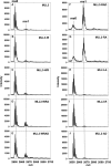
RbBP5/Ash2L heterodimer is required for stimulation of MLL3 methyltransferase activity. MALDI-TOF mass spectrometry showing the histone methylation of an unmodified H3 peptide after 6 h for various subcomplexes containing MLL3. A shows activity with the isolated MLL3 SET domain. B–E show complexes containing WDR5 and F–J show complexes lacking WDR5. The positions for unmodified (me0) and mono-methylated H3 peptide (me1) are indicated above the graphs.
FIGURE 3.
FIGURE 3.
MLL3 assembles core complexes with and without WDR5. Upper panels show the size exclusion chromatographs of MLL3-WRAD (A) or of MLL3-RAD (B) passed over a Superdex 200 column (GE Healthcare). The void volume (45 mls) is indicated on the graphs by a dashed line, and the black bars represent the part of the elution profiles analyzed by SDS-PAGE. The lower panels show Coomassie Blue-stained SDS-PAGE analysis of fractions collected from under the peaks (black bars). Lane 1 of each gel shows the input sample. Diffusion-free sedimentation coefficient distributions (c(s)) were derived from SV-AUC experiments of the MLL3-WRAD (C), MLL3-RAD (D), or RAD (E) complexes at various concentrations.
FIGURE 4.
FIGURE 4.
Win motif is required for the interaction of MLL3 and WDR5. A, comparison of the c(s) distributions for the isolated MLL3WT (solid black line) and MLL3R4710A variant (dashed line) SET domains run at the same concentration. B, c(s) distribution of full-length wild type WDR5. C, comparison of c(s) distributions for the interaction of the wild type (solid black line) or the R4710A variant (dashed line) MLL3 SET domain with WDR5.
FIGURE 5.
FIGURE 5.
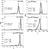
Win motif contributes to the stabilization of WDR5 within the core complex. A and B, comparison of c(s) distributions of the wild type (solid black line) or the R4710A variant (dashed line) MLL3 SET domain assembled with the RAD (A) or WRAD (B) subcomplex. C, comparison of the c(s) distributions of the R4710A variant MLL3 SET domain assembled with WRAD at different concentrations. D and E, comparison of weighted c(s) distributions (c(s)*) (Bayesian analysis) of the wild type (D) or R4710A variant (E) MLL3 SET domain assembled with WRAD.
FIGURE 6.
FIGURE 6.
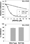
WDR5 partially inhibits the activity of the MLL3 core complex. A, activity assays of the MLL3WT-RAD (filled circles) or MLL3R4710A-RAD (open circles) complexes with increasing amounts of WDR5. Each point represents the mean percentage of total integrated area for each sample in MALDI-TOF reactions after 30 min. Error bars represent ± S.E. from triplicate measurements. The data are normalized to the amount of H3K4me1 activity that either the MLL3WT-RAD or the MLL3R4710A-RAD complexes catalyzed at 30 min when no WDR5 was present. Inhibition curves are fit as described under the “Experimental Procedures.” B, graph indicates that the MLL3-RAD complexes assembled with the wild type or the R4710A variant catalyze the same amount of H3K4 monomethylation (H3K4me1) after a 30-min reaction as determined by MALDI-TOF assays. Error bars represent ± S.E. from triplicate experiments.
FIGURE 7.
FIGURE 7.
MLL3 core complex utilizes two active sites. A, UV light was used to cross-link [3H]AdoMet to wild type or N4848A variant MLL3 SET domains in isolation or assembled with WRAD/RAD. The upper panel shows a Coomassie Blue-stained SDS-polyacrylamide gel, and the lower panel shows the [3H]methyl incorporation after a 72-h exposure to film by fluorography. B, comparison of methyltransferase activities among the wild type or the N4848A variant MLL3 SET domain alone or when assembled with WRAD/RAD. The upper panel shows a Coomassie Blue-stained SDS-polyacrylamide gel, and the lower panels show [3H]methyl incorporation after 4, 24, 48, or 96 h by fluorography. C–E, the c(s) analysis compares the wild type (solid black lines) or the N4848A variant (dotted lines) of the isolated MLL3 SET domain (C), the MLL3-RAD complex (D), or the MLL3-WRAD complex (E).
FIGURE 8.
FIGURE 8.
Biological SAXS characterization of MLL3 core complexes. A, D, and G, Guinier plots of the data showing a linear fit that satisfies qRg of ≤1.3, where Rg is the radius of gyration, and q (momentum transfer) = 4πsin(θ)/λ, where 2θ is the scattering angle, and λ is the x-ray wavelength. The data are plotted as the natural log of the scattering intensity (I) as a function of the square of momentum transfer (q). B, E, H, Plots of the pair distance distribution function, P(r), for MLL3 (B), MLL3-RAD (E), and MLL3-WRAD (H). The maximum intraparticle distance (Dmax) for each plot is reported in Table 3. C, F, and I, plots of the fit of the simulated scattering from the calculated envelopes overlaid with the experimental scattering data. The data are plotted as the log of the intensity (I) as a function of q. The q range used for structural determination is determined such that qmaxRg/8.
FIGURE 9.
FIGURE 9.
Solution structures of MLL3 core complexes. The ab initio solution structures of MLL3(4690–4911) (A), the MLL3-RAD complex (B), and the MLL3-WRAD complex (C) were contoured at 20 Å. The final models represent the refined average model from 9 to 10 individual models generated using programs from the ATSAS package (56) as described under “Experimental Procedures.” A, MLL3 homology model was generated using Modeler (40) and was fit into the MLL3 envelope. D, predicted solution scattering profile of the MLL3 homology model from the FoxS server overlaid with the experimental MLL3 scattering data. E, overlay of the normalized P(r) distributions from the MLL3-WRAD (solid line) and MLL3-RAD (dotted line) complexes. F, rigid body alignment of the MLL3-RAD and MLL3-WRAD envelopes. The WDR5 crystal structure (PDB code 2H14 (49)) is fit into a region of the MLL3-WRAD structure that is missing from the MLL3-RAD structure in the alignment.

References

    1. Strahl B. D., Ohba R., Cook R. G., and Allis C. D. (1999) Methylation of histone H3 at lysine 4 is highly conserved and correlates with transcriptionally active nuclei in Tetrahymena. Proc. Natl. Acad. Sci. U.S.A. 96, 14967–14972 - PMC - PubMed
    1. Chi P., Allis C. D., and Wang G. G. (2010) Covalent histone modifications–miswritten, misinterpreted and mis-erased in human cancers. Nat. Rev. Cancer 10, 457–469 - PMC - PubMed
    1. Milne T. A., Briggs S. D., Brock H. W., Martin M. E., Gibbs D., Allis C. D., and Hess J. L. (2002) MLL targets SET domain methyltransferase activity to Hox gene promoters. Mol. Cell 10, 1107–1117 - PubMed
    1. Nakamura T., Mori T., Tada S., Krajewski W., Rozovskaia T., Wassell R., Dubois G., Mazo A., Croce C. M., and Canaani E. (2002) ALL-1 is a histone methyltransferase that assembles a supercomplex of proteins involved in transcriptional regulation. Mol. Cell 10, 1119–1128 - PubMed
    1. Dou Y., Milne T. A., Tackett A. J., Smith E. R., Fukuda A., Wysocka J., Allis C. D., Chait B. T., Hess J. L., and Roeder R. G. (2005) Physical association and coordinate function of the H3 K4 methyltransferase MLL1 and the H4 K16 acetyltransferase MOF. Cell 121, 873–885 - PubMed

Publication types

MeSH terms

Substances