. 2015 Oct 1;195(7):3382-9.
doi: 10.4049/jimmunol.1500937. Epub 2015 Aug 31.

Retinal Pigment Epithelial Cells Mitigate the Effects of Complement Attack by Endocytosis of C5b-9

Affiliations

Retinal Pigment Epithelial Cells Mitigate the Effects of Complement Attack by Endocytosis of C5b-9

Apostolos Georgiannakis et al. J Immunol. .

Abstract

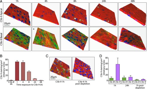
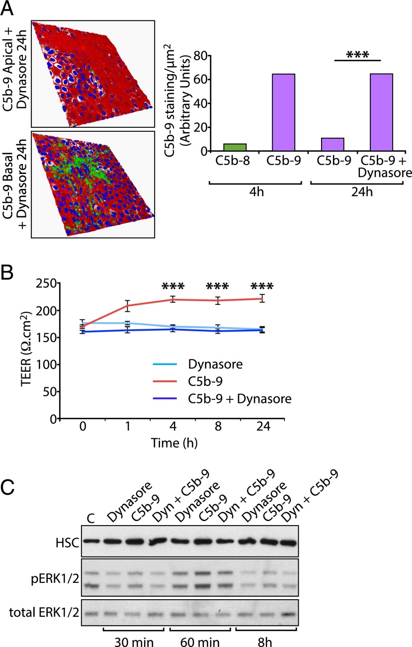
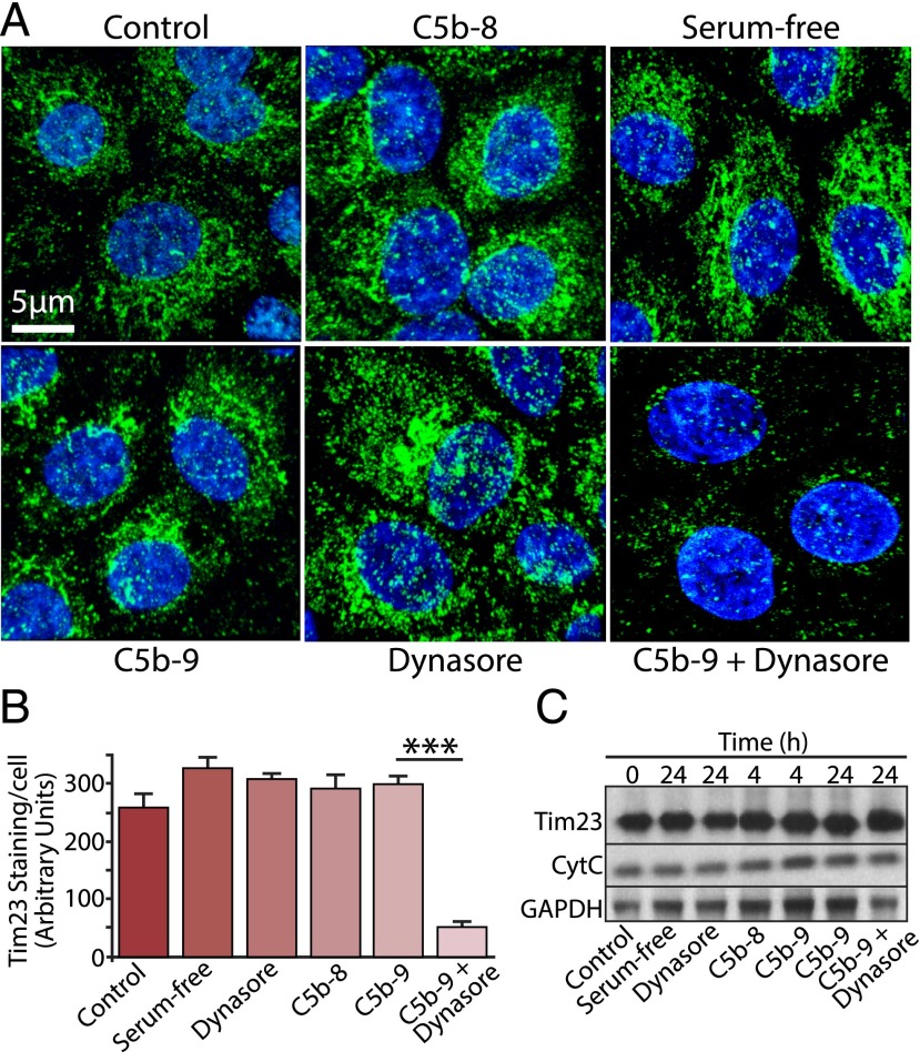
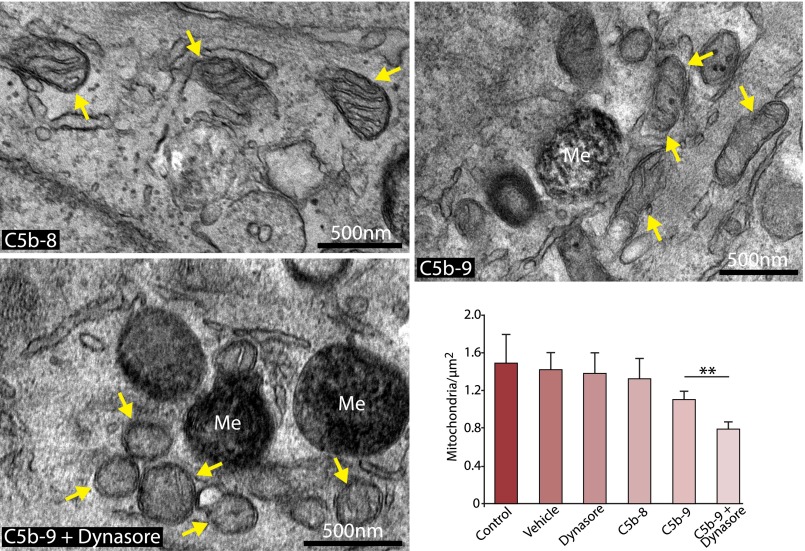
Retinal pigment epithelial (RPE) cell death is a hallmark of age-related macular degeneration. The alternative pathway of complement activation is strongly implicated in RPE cell dysfunction and loss in age-related macular degeneration; therefore, it is critical that RPE cells use molecular strategies to mitigate the potentially harmful effects of complement attack. We show that the terminal complement complex C5b-9 assembles rapidly on the basal surface of cultured primary porcine RPE cells but disappears over 48 h without any discernable adverse effects on the cells. However, in the presence of the dynamin inhibitor dynasore, C5b-9 was almost completely retained at the cell surface, suggesting that, under normal circumstances, it is eliminated via the endocytic pathway. In support of this idea, we observed that C5b-9 colocalizes with the early endosome marker EEA1 and that, in the presence of protease inhibitors, it can be detected in lysosomes. Preventing the endocytosis of C5b-9 by RPE cells led to structural defects in mitochondrial morphology consistent with cell stress. We conclude that RPE cells use the endocytic pathway to prevent the accumulation of C5b-9 on the cell surface and that processing and destruction of C5b-9 by this route are essential for RPE cell survival.


Figures

FIGURE 1.
FIGURE 1.
Accumulation and elimination of C5b-9 on the basal RPE cell surface. (A) Porcine RPE cells were cultured on Transwells and incubated for the periods indicated with DMEM in the apical chamber, as well as with DMEM containing C5b-6, C7, C8, and C9 in the basal chamber. C5b-9 (green), F-actin (red), and DAPI (blue) were visualized by confocal microscopy in full-thickness Z-stacks, of which the apical and basal cell surfaces are shown. *A 3D view is provided in Supplemental Video 1. (B) Quantitation of C5b-9 staining is presented as the average fluorescence intensity of 12 full-depth fields of view from three independent experiments. Omission of C9 from the complement protein mix provided the control C5b-8 sample, which was used in this experiment and elsewhere in this study to demonstrate the specificity of the Ab for C5b-9. (C) RPE cell monolayers were incubated for 1 h as in (A) (left panel) or with complement-containing medium that had been in contact with RPE cells for 24 h (right panel). Monolayers were processed for imaging as in (A). (D) RPE cell monolayers were treated as in (A) for 1 and 24 h, after which the basal DMEM containing the complement protein mix was applied to a new Transwell for 1 h to generate the 1-h postdepletion data. The results show that basal medium taken after 24 h exposure to RPE cells does not support de novo C5b-9 formation. Data were generated from four separate fields of view from three independent experiments and are expressed as mean ± SEM.
FIGURE 2.
FIGURE 2.
Endocytosis is required for the elimination of surface-associated C5b-9. (A) RPE cell monolayers were cultured on Transwells for 24 h in the presence or absence of 200 μM Dynasore, with DMEM containing the complement protein mix in the basal chamber. Cells were processed for imaging as in Fig. 1A. Quantitative analysis shows the expected accumulation of C5b-9 at 4 h and subsequent decline at 24 h. However, in the presence of Dynasore, the level of C5b-9 staining at 24 h was significantly higher and had not decreased significantly from the value at 4 h. (B) To assess the integrity of the monolayer during prolonged exposure to C5b-9 and Dynasore, TEER was measured during the 24-h experimental period. C5b-9 elicited a small, but significant, increase in TEER during this period that was abolished in the presence of Dynasore. Dynasore alone had no effect on TEER. Data are expressed as mean ± SEM [n = 12 fields of view from three independent experiments (A) or 12 wells/condition (B)]. (C) To examine ERK1/2 activation, RPE cells were incubated with 200 μg/ml of Dynasore alone, C5b-9 alone, or the two in combination. Cells were extracted at the times indicated, and whole-cell lysates were analyzed by SDS-PAGE and Western blotting using Abs against p-ERK1/2, total ERK1/2, and heat shock protein 70-kDa chaperone as the loading control. Protein bands were visualized by ECL. ***p < 0.001.
FIGURE 3.
FIGURE 3.
Clearance of C5b-9 via the endocytic pathway. (A) RPE cell monolayers were cultured on Transwells for 4 h in the presence of DMEM containing the complement protein mix and then fixed and immunostained for C5b-9 (green), the early endosomal marker EEA1 (red), and nuclei (DAPI; blue). The representative images show puncta of C5b-9 (white arrows), many of which colocalize with EEA1. (B) Cells were treated as above for 8 h and then fixed and immunostained for C5b-9 (green) and the lysosomal marker cathepsin D (red). Under control conditions (-PL) we observed little colocalization of the two markers. (C) Using the same conditions as in (B) but maintaining the cells in the presence of pepstatin A and leupeptin (+PL), we observed partial colocalization of C5b-9 (white arrows) with cathepsin D, consistent with its trafficking to the lysosome. Note that the variability in DAPI staining is due to the position of the confocal slice relative to the nucleus. (D) Bar graph shows the relative proportion of C5b-9 staining that colocalized with cathepsin D at 4 and 8 h, in the presence and absence of pepstatin A and leupeptin. The numbers were calculated by counting the yellow pixels (corresponding to C5b-9 and cathepsin D colocalization) and dividing these values by the number of yellow plus green (C5b-9) pixels. Data are expressed as mean ± SEM (n = 5 images/experiment from three independent experiments). In the presence of the protease inhibitors there was a significant (**p < 0.05) increase in the colocalization of C5b-9 and cathepsin D from 4 to 8 h and likewise when comparing the control sample at 8 h with the cells treated with the inhibitors (***p < 0.001).
FIGURE 4.
FIGURE 4.
Persistent exposure to C5b-9 leads to mitochondrial perturbation. (A) RPE cells were cultured on Transwells for 24 h and exposed to a variety of experimental conditions, as indicated in the figure. Cells were fixed and immunostained for the mitochondrial marker Tim23 (green) and DAPI (blue). (B) Data are expressed as mean ± SEM (n = 4 images/experiment from three independent experiments). Quantitative analysis of Tim23 staining revealed a significant reduction (***p < 0.001) in staining intensity in cells treated with C5b-9 and Dynasore versus C5b-9 or Dynasore alone. (C) Whole-cell lysates were prepared from RPE cells cultured under the same set of conditions as above for the times indicated and Western blotted for Tim23, cytochrome C (CytC), and GAPDH as a control. No discernable difference was noted in the band intensities for either mitochondrial protein, under any of the different conditions.
FIGURE 5.
FIGURE 5.
Ultrastructural defects in mitochondria exposed to persistent C5b-9. The images show representative transmission electron micrographs of RPE cells that were cultured in Transwells in the presence of C5b-8 (control), C5b-9, and C5b-9+Dynasore for 24 h. Mitochondria are highlighted by arrows. The images show that, in the presence of C5b-9+Dynasore, mitochondria are smaller and rounder than in the other conditions. The bar graph shows a quantitative enumeration of mitochondria number as a function of area, with cells treated with C5b-9+Dynasore exhibiting significantly fewer mitochondria than cells treated with C5b-9 alone. Data are expressed as mean ± SEM (n = 5 images/experiment from three independent experiments). **p < 0.05. Me, melanosome.

Similar articles

Cited by

References

    1. Klein R., Cruickshanks K. J., Nash S. D., Krantz E. M., Nieto F. J., Huang G. H., Pankow J. S., Klein B. E. 2010. The prevalence of age-related macular degeneration and associated risk factors. Arch. Ophthalmol. 128: 750–758. - PMC - PubMed
    1. Mettu P. S., Wielgus A. R., Ong S. S., Cousins S. W. 2012. Retinal pigment epithelium response to oxidant injury in the pathogenesis of early age-related macular degeneration. Mol. Aspects Med. 33: 376–398. - PubMed
    1. Sarks S. H. 1976. Ageing and degeneration in the macular region: a clinico-pathological study. Br. J. Ophthalmol. 60: 324–341. - PMC - PubMed
    1. Bird A. C. 2010. Therapeutic targets in age-related macular disease. J. Clin. Invest. 120: 3033–3041. - PMC - PubMed
    1. Johnson L. V., Ozaki S., Staples M. K., Erickson P. A., Anderson D. H. 2000. A potential role for immune complex pathogenesis in drusen formation. Exp. Eye Res. 70: 441–449. - PubMed

Publication types

MeSH terms

Substances

Supplementary concepts

LinkOut - more resources