. 2015 Oct;169(2):1155-67.
doi: 10.1104/pp.15.00214. Epub 2015 Aug 31.

PLASTID MOVEMENT IMPAIRED1 and PLASTID MOVEMENT IMPAIRED1-RELATED1 Mediate Photorelocation Movements of Both Chloroplasts and Nuclei

Affiliations

PLASTID MOVEMENT IMPAIRED1 and PLASTID MOVEMENT IMPAIRED1-RELATED1 Mediate Photorelocation Movements of Both Chloroplasts and Nuclei

Noriyuki Suetsugu et al. Plant Physiol. 2015 Oct.

Abstract

Organelle movement and positioning play important roles in fundamental cellular activities and adaptive responses to environmental stress in plants. To optimize photosynthetic light utilization, chloroplasts move toward weak blue light (the accumulation response) and escape from strong blue light (the avoidance response). Nuclei also move in response to strong blue light by utilizing the light-induced movement of attached plastids in leaf cells. Blue light receptor phototropins and several factors for chloroplast photorelocation movement have been identified through molecular genetic analysis of Arabidopsis (Arabidopsis thaliana). PLASTID MOVEMENT IMPAIRED1 (PMI1) is a plant-specific C2-domain protein that is required for efficient chloroplast photorelocation movement. There are two PLASTID MOVEMENT IMPAIRED1-RELATED (PMIR) genes, PMIR1 and PMIR2, in the Arabidopsis genome. However, the mechanism in which PMI1 regulates chloroplast and nuclear photorelocation movements and the involvement of PMIR1 and PMIR2 in these organelle movements remained unknown. Here, we analyzed chloroplast and nuclear photorelocation movements in mutant lines of PMI1, PMIR1, and PMIR2. In mesophyll cells, the pmi1 single mutant showed severe defects in both chloroplast and nuclear photorelocation movements resulting from the impaired regulation of chloroplast-actin filaments. In pavement cells, pmi1 mutant plants were partially defective in both plastid and nuclear photorelocation movements, but pmi1pmir1 and pmi1pmir1pmir2 mutant lines lacked the blue light-induced movement responses of plastids and nuclei completely. These results indicated that PMI1 is essential for chloroplast and nuclear photorelocation movements in mesophyll cells and that both PMI1 and PMIR1 are indispensable for photorelocation movements of plastids and thus, nuclei in pavement cells.


Figures

Figure 1.
Figure 1.
Gene structure of PMI1, PMIR1, and PMIR2 and chloroplast photorelocation movement in mesophyll cells of pmi1 and pmir1 pmir2 mutants. A, Gene structure and mutation sites of PMI1, PMIR1, and PMIR2 genes. Rectangles indicate exons (gray rectangles indicate 5′ or 3′ untranslated region), and intervening bars indicate introns. The gray bar in PMI1 shows the promoter region used in PMI1pro::PMI1-GFP. LB, Left border of T-DNA. B, Changes in leaf transmittance caused by chloroplast photorelocation movement. After transmittance measurement started, dark-adapted samples were kept in darkness for an additional 10 min. Then, samples were sequentially irradiated with continuous blue light at 3, 20, and 50 µmol m−2 s−1 for 60, 40, and 40 min indicated by white, sky blue, and blue arrows, respectively. Light was turned off at 150 min (black arrow). Mean values from three independent experiments are shown. Error bars indicate ses. C, Changes in leaf transmittance rates from 2 to 6 min after changes in light fluence rate (3, 20, and 50 µmol m−2 s−1) are indicated as percentage transmittance change over 1 min. Mean values from three independent experiments are shown. Error bars indicate ses. WT, Wild type.
Figure 2.
Figure 2.
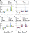
Changes in leaf transmittance rates in mesophyll cells of mutants crossed between pmi1 and phot, jac1, web1, or pmi2. Changes in leaf transmittance rates from 2 to 6 min after changes in light fluence rate (3, 20, and 50 µmol m−2 s−1). A, Genetic interaction between PMI1 and PHOT genes. B, Genetic interaction between PMI1 and JAC1, WEB1, and PMI2 (and PMI15) genes. C, Genetic interaction between PMI1, JAC1, and WEB1 genes. D, Genetic interaction between PMI1, JAC1, and PMI2 (and PMI15) genes. For details, see Figure 1C. Mean values from three independent experiments are shown. Error bars indicate ses. WT, Wild type.
Figure 3.
Figure 3.
Subcellular localization of PMI1 and fractionation of protein factors regulating chloroplast movement in pmi1. A, Subcellular localization of PMI1-GFP. Transverse sections of pavement cells and mesophyll cells were observed under a confocal laser-scanning microscope. Image is false colored to indicate fluorescence of GFP (green) and chlorophyll (red). Arrows indicate PMI1-GFP fluorescence in the cytoplasm. B, Immunoblot analysis of PHOT1, PHOT2, JAC1, CHUP1, and KAC proteins in various mutants. Total protein extracts (T) were fractionated into soluble (S) and microsomal (M) fractions by ultracentrifugation (100,000g for 30 min at 4°C). Immunoblotting was performed using indicated antisera (Suetsugu et al., 2010b). Numbers on the left indicate the molecular weight of protein markers in the far left lanes. Arrows indicate deduced full-length bands of indicated proteins. The small arrow indicates the phot1 protein band recognized by phot2 antisera. WT, Wild type.
Figure 4.
Figure 4.
Observation of cp-actin filaments on moving chloroplasts in mesophyll cells of wild-type and pmi1 cells. A and B, Time-lapse images of reorganization of cp-actin filaments in wild-type (A) and pmi1 (B) cells during chloroplast movement in response to strong blue light (BL). Actin filaments were probed with GFP-mouse talin fusion protein (green). Blue broken lines indicate the BL-irradiated area. Irradiation times (minutes-seconds) are shown at the top left corner. Note that cp-actin filaments rapidly reorganized on the rims of moving chloroplasts (numbers 1–6). White arrows indicate rapid disappearance of cp-actin filaments from the rear region of moving chloroplasts; yellow arrows indicate reappearance of cp-actin filaments in the front region of moving chloroplasts. For the full time-lapse series, see Supplemental Movie S1. Bar = 10 µm.
Figure 5.
Figure 5.
Reorganizations of cp-actin filaments in mesophyll cells under different light conditions. A, Light-dependent reorganization of cp-actin filaments. Cells of wild-type (WT) and pmi1 leaves were irradiated with serial scans of a 458-nm laser for 30 s (blue light [BL] 30 s) and then incubated in the dark for 4 min (dark [D] 4 min). Next, 3-min serial scans with 458- and 488-nm lasers (BL 3 min) were carried out to induce disappearance of cp-actin filaments. Finally, cells were incubated in the dark for 4 min (D 4 min). Images are false colored to show GFP (green) and chlorophyll (red) fluorescence. Note that cp-actin filaments disappeared after BL irradiation and reappeared after 4 min of adaptation in the dark in both the wild type and pmi1. Bar = 5 µm. B, BL-induced disappearance of cp-actin filaments in wild-type and pmi1 mutant cells. Fluorescence intensities of cp-actin filaments were measured at chloroplast edges in wild-type and pmi1 mutant cells, representing changes in the amount of cp-actin filaments during BL irradiation for 3 min after the 4-min dark adaption. Values are mean ± sd (n = 5 squares) in arbitrary units. C and D, Effect of 488- (C) and 516-nm (D) imaging lasers on avoidance response in pmi1 mutant cells. Time-lapse images were collected at approximately 30-s intervals with two different imaging lasers (488 and 516 nm) for 15 min and 8 s. The blue rectangular regions (region of interest [roi], 10 × 20 µm) were irradiated with a stimulating laser (458 nm) during intervals between the image acquisitions of chlorophyll fluorescence images with the imaging lasers. Chlorophyll fluorescence is false colored in red. Right shows moving paths of individual chloroplasts (a–d). For the full time-lapse series, see Supplemental Movie S2. Bars = 10 µm.
Figure 6.
Figure 6.
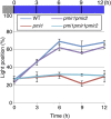
Distinct roles of PMI1 and PMIRs on nuclear photorelocation movement in mesophyll cells. Time-course analysis of nuclear avoidance response in mesophyll cells of wild-type (WT), pmi1, pmir1pmir2 double-mutant, and their triple-mutant plants. Nuclear avoidance response was induced by strong blue light (50 µmol m−2 s−1). The percentage of cells in which the nucleus was in the light position is depicted in mean ± sd. Each data point was obtained from five leaves; 100 cells were observed in each leaf.
Figure 7.
Figure 7.
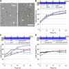
Distinct roles of PMI1 and PMIRs on nuclear photorelocation movement in pavement cells. A, Representative images showing dark position (left) and light position (right) of nuclei under the strong blue light (BL) in pavement cells of wild-type Arabidopsis. Bar = 25 µm. B to D, Time-course analysis of nuclear avoidance response in pavement cells of wild-type (WT), pmi1, pmir1, pmir2 single, and their double- and triple-mutant plants. The other details are the same as in Figure 6.

References

    1. Castresana J. (2000) Selection of conserved blocks from multiple alignments for their use in phylogenetic analysis. Mol Biol Evol 17: 540–552 - PubMed
    1. Cazzaniga S, Dall’ Osto L, Kong SG, Wada M, Bassi R (2013) Interaction between avoidance of photon absorption, excess energy dissipation and zeaxanthin synthesis against photooxidative stress in Arabidopsis. Plant J 76: 568–579 - PubMed
    1. Chevenet F, Brun C, Bañuls AL, Jacq B, Christen R (2006) TreeDyn: towards dynamic graphics and annotations for analyses of trees. BMC Bioinformatics 7: 439. - PMC - PubMed
    1. Cutler SR, Ehrhardt DW, Griffitts JS, Somerville CR (2000) Random GFP:cDNA fusions enable visualization of subcellular structures in cells of Arabidopsis at a high frequency. Proc Natl Acad Sci USA 97: 3718–3723 - PMC - PubMed
    1. Davis PA, Hangarter RP (2012) Chloroplast movement provides photoprotection to plants by redistributing PSII damage within leaves. Photosynth Res 112: 153–161 - PubMed

Publication types

MeSH terms

Substances