. 2015 Sep 15;112(37):E5169-78.
doi: 10.1073/pnas.1505512112. Epub 2015 Aug 31.

Differential control of Yorkie activity by LKB1/AMPK and the Hippo/Warts cascade in the central nervous system

Affiliations

Differential control of Yorkie activity by LKB1/AMPK and the Hippo/Warts cascade in the central nervous system

Ieva Gailite et al. Proc Natl Acad Sci U S A. .

Abstract

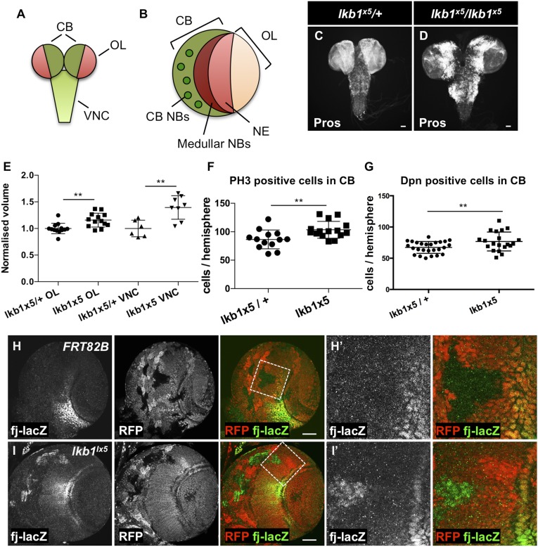
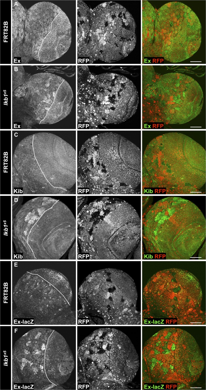
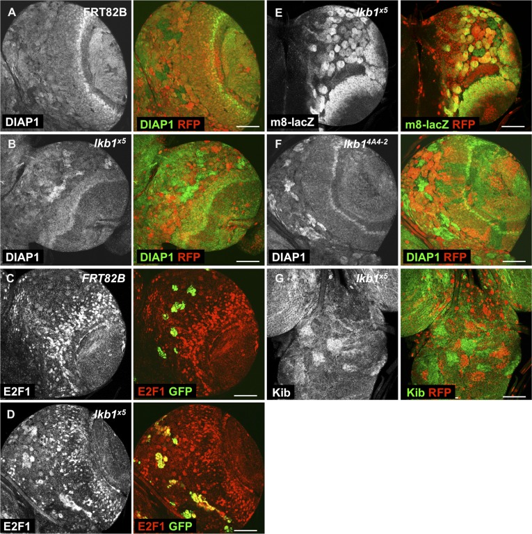
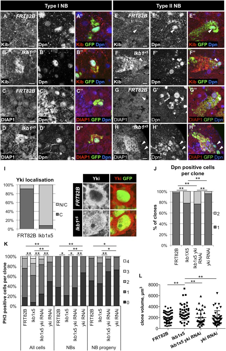
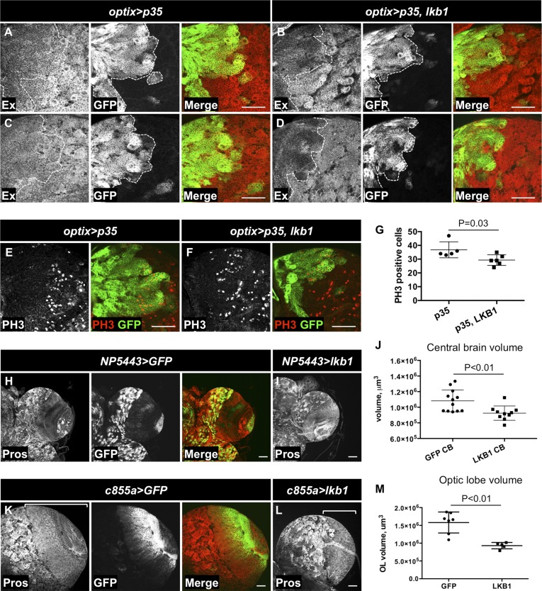
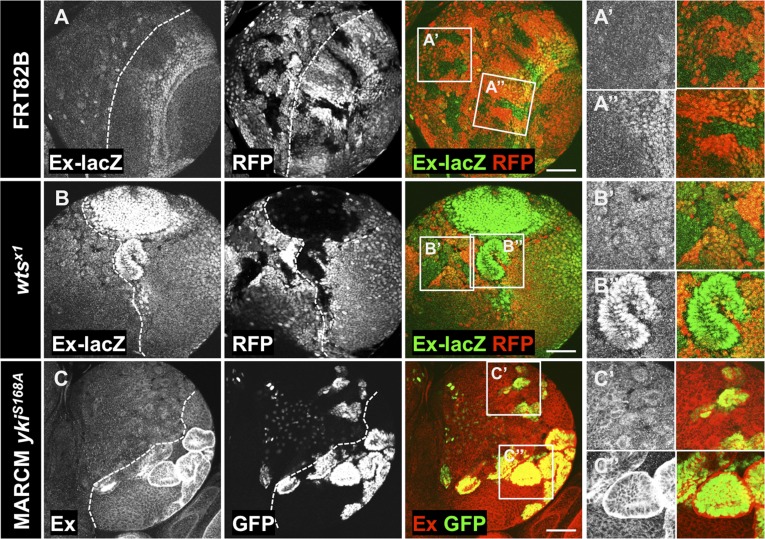
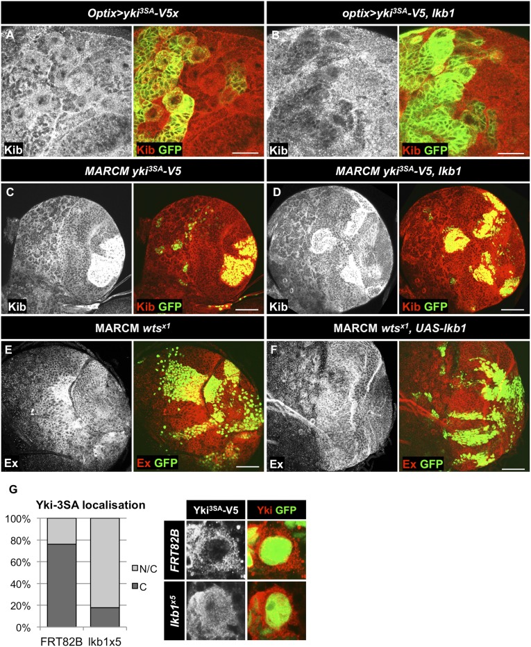
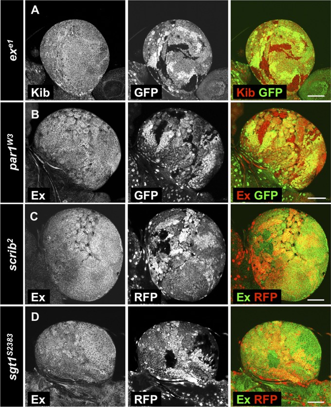
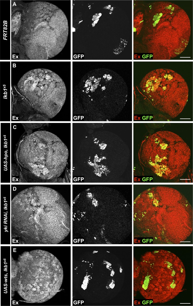
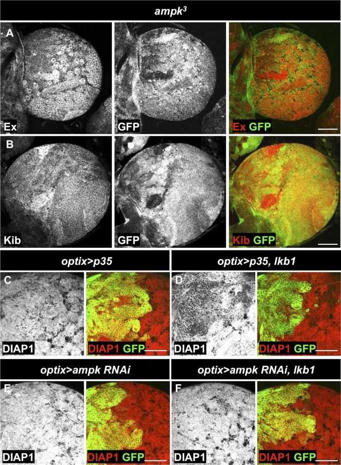
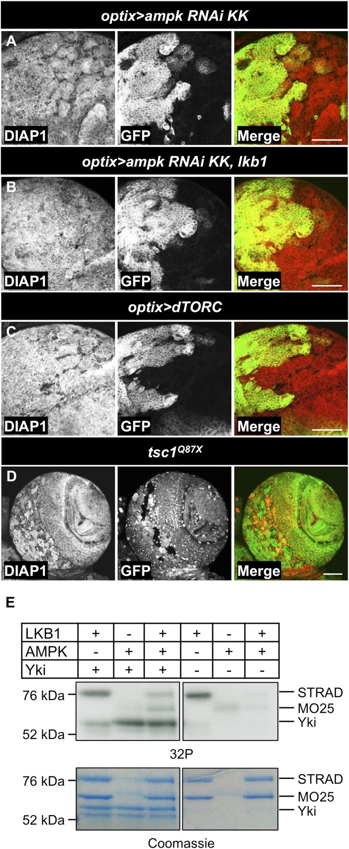
The Hippo (Hpo) pathway is a highly conserved tumor suppressor network that restricts developmental tissue growth and regulates stem cell proliferation and differentiation. At the heart of the Hpo pathway is the progrowth transcriptional coactivator Yorkie [Yki-Yes-activated protein (YAP)/transcriptional coactivator with PDZ-binding motif (TAZ) in mammals]. Yki activity is restricted through phosphorylation by the Hpo/Warts core kinase cascade, but increasing evidence indicates that core kinase-independent modes of regulation also play an important role. Here, we examine Yki regulation in the Drosophila larval central nervous system and uncover a Hpo/Warts-independent function for the tumor suppressor kinase liver kinase B1 (LKB1) and its downstream effector, the energy sensor AMP-activated protein kinase (AMPK), in repressing Yki activity in the central brain/ventral nerve cord. Although the Hpo/Warts core cascade restrains Yki in the optic lobe, it is dispensable for Yki target gene repression in the late larval central brain/ventral nerve cord. Thus, we demonstrate a dramatically different wiring of Hpo signaling in neighboring cell populations of distinct developmental origins in the central nervous system.

Keywords: Hippo; development; growth control; neuroblast; stem cells.


Conflict of interest statement

The authors declare no conflict of interest.

Figures

Fig. 1.
Fig. 1.
Loss of lkb1 function causes overgrowth of the larval brain. (A) Schematic representation of the compartments of the larval CNS. (B) Schematic representation of the third instar larval brain hemisphere. CB, central brain; NB, neuroblasts; NE, neuroepithelium; OL, optic lobe; VNC, ventral nerve cord. (C and D) CNS morphology of lkb1x5/+ (C) and lkb1x5/lkb1x5 (D) white prepupae. Pros staining labels neuronal cells. (E) Volume of lkb1x5 homozygous VNCs (n = 8) and OLs (n = 13) is increased in comparison with heterozygous lkb1x5/+ VNCs (n = 6) and OLs (n = 13). Brains were dissected from white prepupae. (F) lkb1x5 homozygous third instar larval brains (n = 15) have increased PH3-positive cell numbers in the CB in comparison with lkb1x5/+ controls (n = 13). (G) lkb1x5 homozygous third instar larval brains (n = 20) show an increase in Dpn-positive cell numbers in the CB in comparison with lkb1x5/+ controls (n = 26). **P < 0.01. Statistical significance was analyzed using Student’s t test. (H and I′) Heat shock (hs) FLP/FRT-generated lkb1x5 clones have increased fj-lacZ levels in the OL (H and H′) in comparison with the control clones (I and I′). Higher magnifications of the boxed areas are shown in H′ and I′. Clones are marked by the absence of the Red Fluorescent Protein (RFP) expression. (Scale bars, 50 μm.)
Fig. 2.
Fig. 2.
lkb1 restricts Yki target expression in the larval CB. Expression levels of the Yki targets Ex (B), Kib (D), and the transcriptional reporter ex-lacZ (F) are elevated in hsFLP/FRT-generated lkb1 mutant clones in the CB of third instar larvae compared with wild-type clones (A, C, and E). Clones are marked by the absence of RFP expression. OL and CB compartments are separated by a line. (Scale bars, 50 μm.)
Fig. S1.
Fig. S1.
lkb1 regulates Yki target expression in the larval CB. (A and B) Levels of DIAP1 protein are increased in hsFLP/FRT-generated lkb1x5 clones (B) but not control clones (A) in the CB. Clones are marked by absence of RFP. (C and D) Levels of E2F1 protein are elevated in lkb1x5 MARCM clones (D), but not control MARCM clones (C), in the CB. Clones are marked by expression of GFP. (E) Expression of the E(spl)m8-lacZ transcriptional reporter is unchanged in lkb1x5 clones. Clones are marked by absence of RFP. (F) Levels of DIAP1 protein are elevated in hsFLP/FRT-generated lkb14A4-2 mutant clones in the CB. Clones are marked by absence of RFP. (G) Increased Kib protein levels in lkb1x5 clones in the VNC. Clones are marked by absence of RFP. (Scale bars, 50 μm.)
Fig. 3.
Fig. 3.
Yki target gene expression in lkb1 mutant NBs and their progeny. Expression of Yki target genes in MARCM clones of type I NBs (A–D′′) and type II NBs (E–H′′). anti-Dpn staining marks type I NBs in A–D′) and type II NBs and mature intermediate neural progenitors in E–H′. Type II NBs are marked by arrowheads in E–H′. Kib (A and E) and DIAP1 (C and G) are uniformly expressed both in NBs and their progeny in control MARCM clones. Protein levels of Kib (B and F) are increased both in type I and type II NBs and their progeny in lkb1x5 MARCM clones. (D and H) In lkb1x5 mutant clones, DIAP1 protein levels are increased in type I NBs and in the type I and type II NB progeny, but not in the type II NBs (H). Clones are marked by GFP expression. (Scale bars, 10 μm.) (I) Quantification of Yki localization in NBs of MARCM clones of the indicated genotypes in third instar larval CB. C, cytoplasmic Yki; N/C, cytoplasmic and nuclear Yki. n = 66 (FRT82B), 64 (lkb1x5). (J) Quantification of Dpn-positive cells in type I NB MARCM clones of the indicated genotypes 48 h after clone induction in the CB of third instar larvae. n = 46 (FRT82B and yki RNAi), 57 (lkb1x5), 51 (lkb1x5 yki RNAi). (K) Quantification of PH3-positive cells in MARCM clones of the indicated genotypes in type I NB lineages in the third instar larval VNC. n = 78 (FRT82B), 69 (lkb1x5), 71 (lkb1x5 yki RNAi), 51 (yki RNAi). (L) Quantification of the volume of MARCM clones of the indicated genotypes 72 h after induction in the third instar larval VNC. n = 52 (FRT82B), 42 (lkb1x5), 33 (lkb1x5 yki RNAi), 38 (yki RNAi). *P < 0.05; **P < 0.01. Statistical significance was analyzed using Χ2 test in J and Student’s t test in K and L.
Fig. 4.
Fig. 4.
LKB1 reduces Yki target gene expression and growth in the CB. (A–D) Overexpression of LKB1 under the control of optix-Gal4 in a subset of CB NBs and their progeny reduces protein levels of Kib and Ex. The caspase inhibitor p35 was coexpressed to prevent cell death upon LKB1 over-expression. (E–G) Overexpression of LKB1 reduces the number of PH3-labeled mitotic cells in the optix compartment of the CB. The optix compartment is marked by GFP expression in A–F. (H) Expression pattern of P(GawB)NP5443 in the CB of white prepupae. (I and J) Overexpression of LKB1 in the CB reduces its volume. Pros staining labels neuronal cells in the CB. (K–M) Overexpression of LKB1 in the OL using c855a-GAL4 reduces its volume. The OL compartment in K and L is marked with a bracket. (Scale bars, 50 μm.) Statistical significance was analyzed using a Student’s t test in G, J, and M.
Fig. 5.
Fig. 5.
Wts is dispensable for Yki inhibition in the CB. (A and B) Expression of ex-lacZ transcriptional reporter in control (AA′′) or wtsx1 (BB′′) mutant clones. Loss of wts function increases expression of ex-lacZ transcriptional reporter in the OL (B′′) in comparison with the control clones (A′′), but not in the CB (B′). ex-lacZ expression in the control clones in the CB is shown in (A′). Clones are marked by absence of RFP. MARCM clones overexpressing YkiS168A (CC′′) induce accumulation of Ex protein in the OL (C′′), but not in the CB (C′). Clones are marked by GFP expression. OL and CB areas are separated by a dashed line in A, B, C, and D. Insets show magnifications of the boxed areas. (Scale bars, 50 μm.)
Fig. S2.
Fig. S2.
LKB1 inhibits Yki activity downstream of Wts. (A and B) LKB1 overexpression inhibits Yki target gene expression in the presence of YkiS111A S168A S250A (Yki3SA). (A) Overexpression of Yki3SA alone in the optix compartment does not modify protein levels of Yki target gene Kib. (B) Expression of LKB1 together with Yki3SA in the optix compartment reduces Kib protein levels. The optix compartment is marked by GFP expression. (C and D) LKB1 overexpression is not sufficient to restore Kib levels in Yki3SA-expressing MARCM clones in the optic lobe to wild-type levels. (E) Protein levels of the Yki target Ex are increased in wtsx1 clones generated with MARCM system in the NE of third instar larval brains. (F) Overexpression of LKB1 in wtsx1 clones reduces Ex levels caused by wts loss. Clones are marked by GFP expression. (Scale bars, 20 μm in A and B and 50 μm in C and F.) (G) Quantification of Yki3SA-V5 localization in NBs of MARCM clones of the indicated genotypes in third instar larval CB. C, cytoplasmic Yki; N/C, cytoplasmic and nuclear Yki. n = 67 (FRT82B), 51 (lkb1x5).
Fig. S3.
Fig. S3.
Known polarity and growth regulators do not regulate Yki target genes in the CB. (A) Protein levels of the Yki target gene Kib are unchanged in exe1 mutant clones. Protein levels of the Yki target Ex are not affected in the mutant clones for par-1w3 (B), scrib2 (C), and sgt1S2383 (D) in third instar larval CBs. Clones are marked by absence of GFP (A and B) or absence of RFP (C and D). (Scale bars, 50 μm.)
Fig. 6.
Fig. 6.
lkb1 regulates Yki target genes downstream of wts. (A–E) MARCM clones in third instar larval CBs. (A) Ex protein levels in control clones. (B) lkb1x5 mutant clones accumulate Ex. (C) Overexpression of UAS-hpo does not rescue up-regulation of Yki targets. Expression of UAS-yki RNAi (D) or UAS-wts (E) rescues Ex accumulation caused by loss of lkb1 function. Clones are marked by GFP expression. (Scale bars, 50 μm.)
Fig. 7.
Fig. 7.
Loss of ampk function up-regulates Yki target genes. (A and B) Protein levels of Kib and Ex are increased in hsFLP/FRT-generated ampk3 clones. Clones are marked by the absence of GFP expression. (C–F) RNAi against ampk rescues inhibition of Yki target gene expression by LKB1. LKB1 overexpression in the optix compartment (D) reduces DIAP1 protein levels in comparison with the control (C). The caspase inhibitor p35 was coexpressed to prevent cell death upon LKB1 over-expression in (C and D). (E) Expression of UAS-ampk RNAiGD736 alone does not modify DIAP1 levels. (F) Expression of UAS-ampk RNAiGD736 together with UAS-lkb1 rescues the reduction of DIAP1 levels. The optix compartment is marked by GFP expression in C–E. (Scale bars, 50 μm.)
Fig. S4.
Fig. S4.
Reduction of AMPK activity rescues LKB1 overexpression phenotypes. (A) Expression of UAS-ampk RNAiKK102684 in the optix compartment does not modify DIAP1 protein levels. (B) Wild-type DIAP1 levels in cells expressing UAS-ampk RNAiKK102684 together with UAS-lkb1. (C) Expression of dTORC in the optix compartment does not affect DIAP1 protein levels. The optix compartment is marked by GFP expression (A–C). (D) DIAP1 protein levels are not changed in hsFLP/FRT-generated tsc1Q87X clones. Clones are marked by absence of RFP expression. (Scale bars, 50 μm.) (E) Human AMPK phosphorylates Yki in vitro. In vitro kinase assays using [γ-32P] ATP were performed with Flag-Yki, affinity purified from Sf9 cells, and human LKB1/MO25/STRAD and AMPK complexes. Protein loading is shown by SimplyBlue SafeStain staining. Presence of AMPK markedly increased Yki phosphorylation.

Similar articles

Cited by

References

    1. Yu FX, Guan KL. The Hippo pathway: Regulators and regulations. Genes Dev. 2013;27(4):355–371. - PMC - PubMed
    1. Lin JI, Poon CL, Harvey KF. The Hippo size control pathway--ever expanding. Sci Signal. 2013;6(259):pe4. - PubMed
    1. Nolo R, Morrison CM, Tao C, Zhang X, Halder G. The bantam microRNA is a target of the hippo tumor-suppressor pathway. Curr Biol. 2006;16(19):1895–1904. - PubMed
    1. Thompson BJ, Cohen SM. The Hippo pathway regulates the bantam microRNA to control cell proliferation and apoptosis in Drosophila. Cell. 2006;126(4):767–774. - PubMed
    1. Huang J, Wu S, Barrera J, Matthews K, Pan D. The Hippo signaling pathway coordinately regulates cell proliferation and apoptosis by inactivating Yorkie, the Drosophila Homolog of YAP. Cell. 2005;122(3):421–434. - PubMed

Publication types

MeSH terms

LinkOut - more resources