. 2015 Oct 6;112(40):E5543-51.
doi: 10.1073/pnas.1510516112. Epub 2015 Aug 31.

Arabidopsis ALIX is required for the endosomal localization of the deubiquitinating enzyme AMSH3

Affiliations

Arabidopsis ALIX is required for the endosomal localization of the deubiquitinating enzyme AMSH3

Kamila Kalinowska et al. Proc Natl Acad Sci U S A. .

Abstract

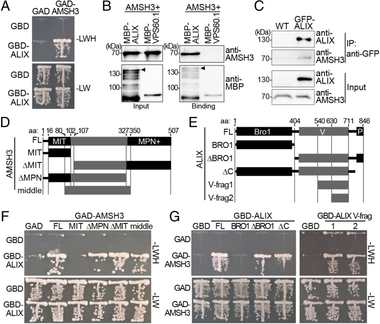
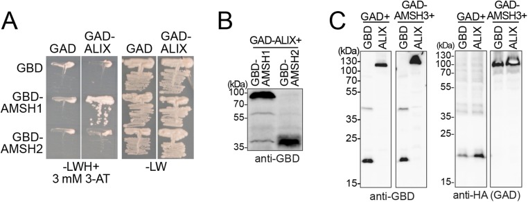
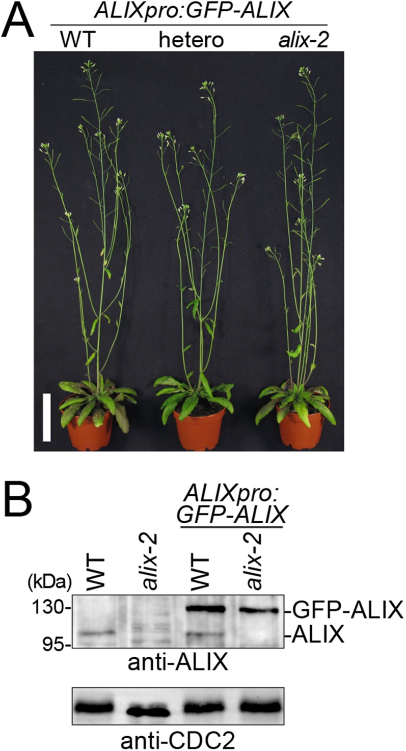
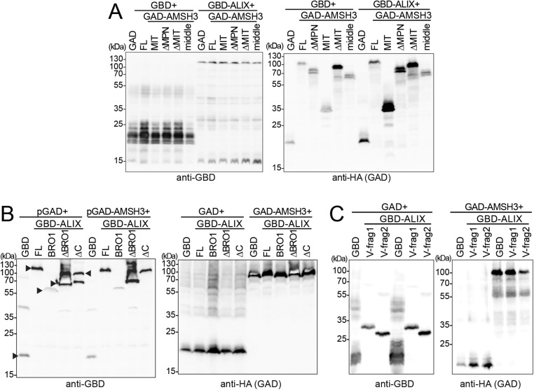
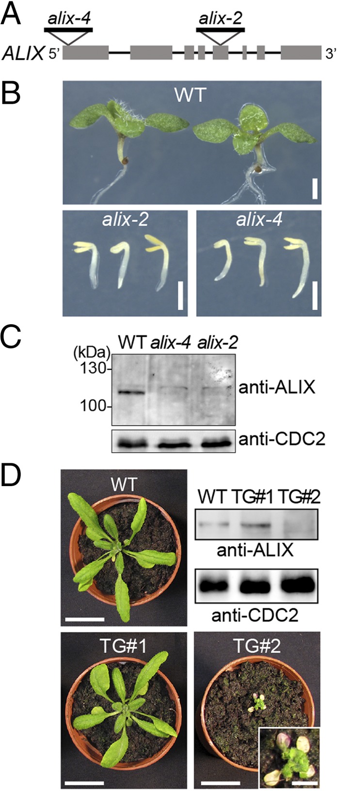
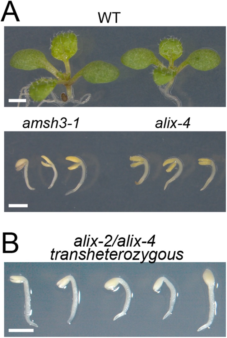
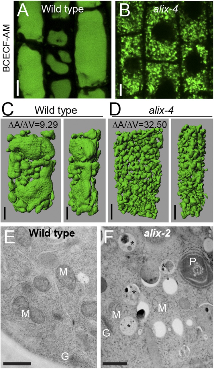
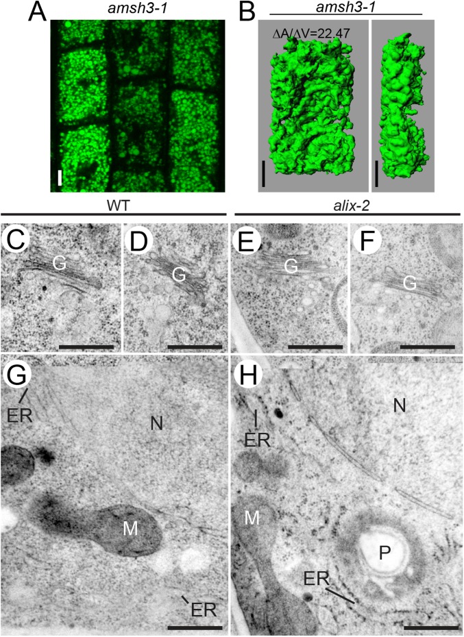
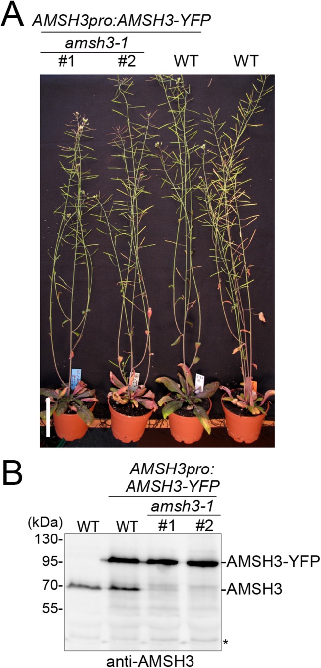
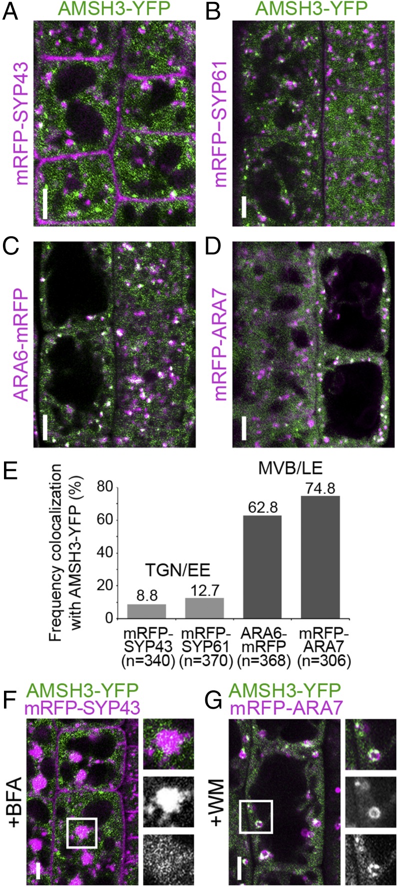
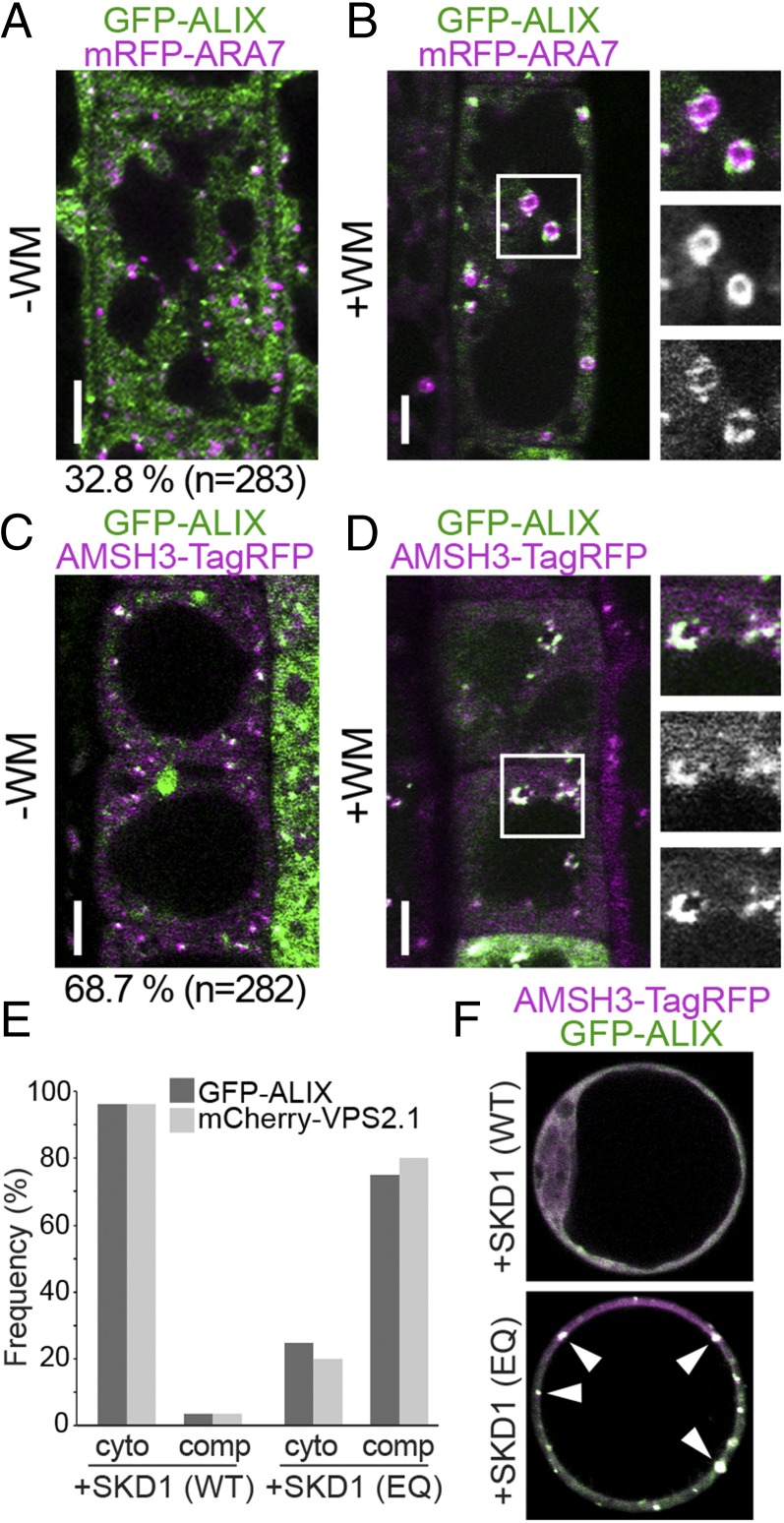
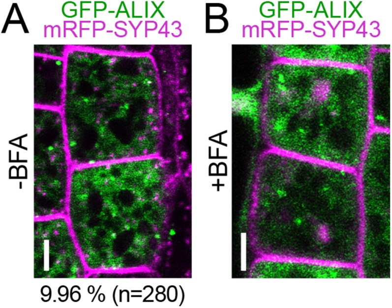
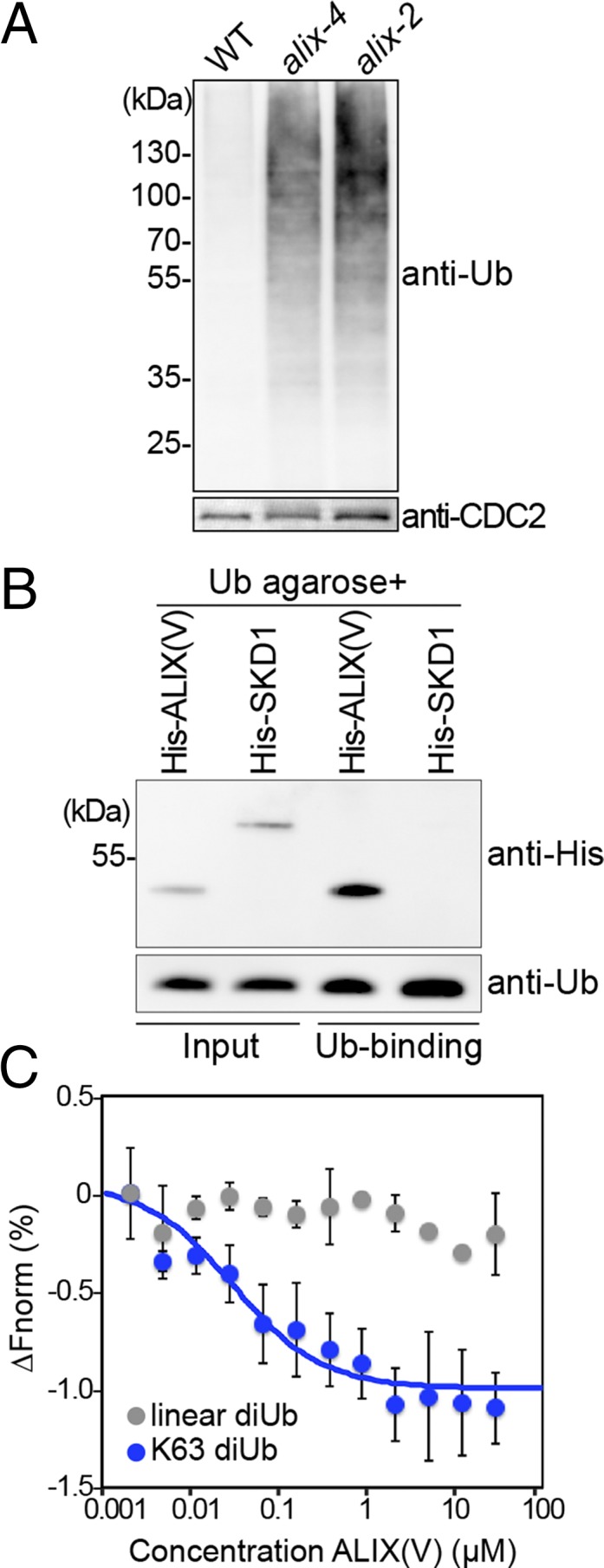
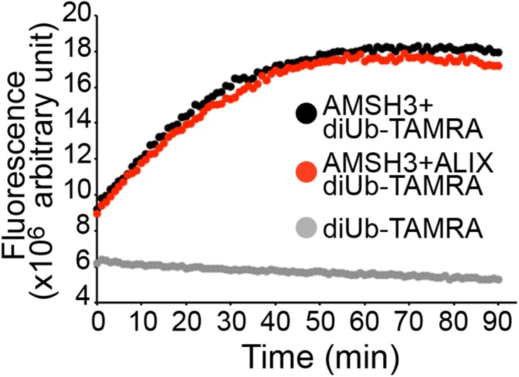
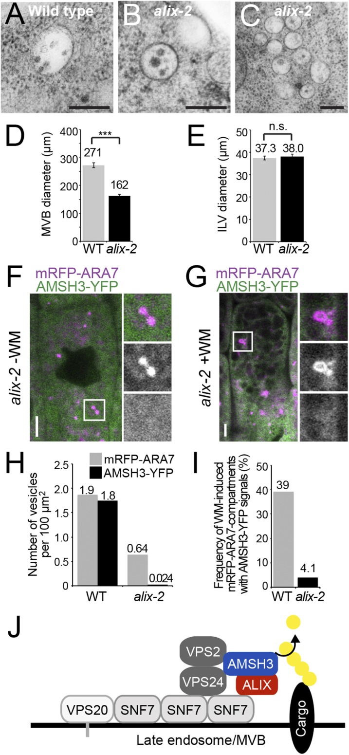
Ubiquitination is a signal for various cellular processes, including for endocytic degradation of plasma membrane cargos. Ubiquitinating as well as deubiquitinating enzymes (DUBs) can regulate these processes by modifying the ubiquitination status of target protein. Although accumulating evidence points to the important regulatory role of DUBs, the molecular basis of their regulation is still not well understood. Associated molecule with the SH3 domain of signal transduction adaptor molecule (STAM) (AMSH) is a conserved metalloprotease DUB in eukaryotes. AMSH proteins interact with components of the endosomal sorting complex required for transport (ESCRT) and are implicated in intracellular trafficking. To investigate how the function of AMSH is regulated at the cellular level, we carried out an interaction screen for the Arabidopsis AMSH proteins and identified the Arabidopsis homolog of apoptosis-linked gene-2 interacting protein X (ALIX) as a protein interacting with AMSH3 in vitro and in vivo. Analysis of alix knockout mutants in Arabidopsis showed that ALIX is essential for plant growth and development and that ALIX is important for the biogenesis of the vacuole and multivesicular bodies (MVBs). Cell biological analysis revealed that ALIX and AMSH3 colocalize on late endosomes. Although ALIX did not stimulate AMSH3 activity in vitro, in the absence of ALIX, AMSH3 localization on endosomes was abolished. Taken together, our data indicate that ALIX could function as an important regulator for AMSH3 function at the late endosomes.

Keywords: Arabidopsis; ESCRT-III; intracellular trafficking; ubiquitin.


Conflict of interest statement

The authors declare no conflict of interest.

Figures

Fig. 1.
Fig. 1.
(A) ALIX interacts with AMSH3 in YTH. GBD and GBD-ALIX were transformed in yeast with either GAD- or GAD-AMSH3. Transformants were tested for their auxotrophic growth on synthetic complete medium lacking leucine and tryptophan (SC-LW) (-LW: SC-Leu-Trp) or synthetic complete medium lacking leucine, tryptophan, and histidine (SC-LWH) (-LWH: SC-Leu-Trp-His) media. (B) ALIX interacts directly with AMSH3 in vitro. Recombinant AMSH3 was incubated with either MBP-ALIX or MBP-VPS60.1 for 1 h at 4 °C and subjected to immunoblot analysis after extensive washing. Anti-AMSH3 and anti-MBP antibodies were used to detect beads-retained material. Arrowheads indicate MBP-ALIX. (C) ALIX and AMSH3 interact in planta. Immunoprecipitation (IP) was performed from total extracts of wild-type or GFP-ALIX-expressing seedlings using anti-GFP immobilized matrix. Immunoprecipitated material was subjected to immunoblot analysis. GFP-ALIX and endogenous AMSH3 were detected with anti-ALIX and anti-AMSH3 antibodies, respectively. (D and E) AMSH3- (D) and ALIX- (E) constructs used for YTH interaction studies in F and G. Bro1, Bro1-domain; FL, full-length; MIT, MIT domain; MPN+, MPN+ domain; P, Proline-rich domain; V, V-domain; V-frag1/2, V-domain fragment 1/2. (F) YTH analysis of AMSH3 domains responsible for the interaction with ALIX. GAD-fused AMSH3 constructs shown in D were cotransformed with either GBD or GBD-ALIX. Transformants were tested for their auxotrophic growth on SC-LW and SC-LWH media. (G) YTH analysis of ALIX domains responsible for the interaction with AMSH3. GBD-fused ALIX constructs shown in E were cotransformed with either GAD or GAD-AMSH3. Transformants were tested for their auxotrophic growth on SC-LW and SC-LWH media.
Fig. S1.
Fig. S1.
(A) ALIX interacts with AMSH1 in a YTH assay. Growth of representative colonies on SC-LW (-LW: SC-Leu-Trp) and SC-LWH (-LWH: SC-Leu-Trp-His) media supplemented with 3 mM 3-AT are shown. (B) Expression of the YTH clones shown in A. Yeast total extracts were prepared from transformants shown in A, and expression of GBD-AMSH1 and GBD-AMSH2 was verified using an anti-GBD antibody. (C) Expression of the YTH clones shown in Fig. 1A. Yeast total extracts were prepared from transformants shown in Fig. 1A and subjected to immunoblotting, using an anti-GBD antibody to detect GBD-fusion proteins and an anti-HA antibody to detect the GAD-HA-fusion proteins.
Fig. S2.
Fig. S2.
Complementation of alix-2 with ALIXpro:GFP-ALIX. (A) ALIXpro:GFP-ALIX complements seedling lethality of alix-2. Wild-type (WT), heterozygous alix-2 (hetero), and homozygous alix-2 plants expressing GFP-ALIX are shown. (Scale bar, 5 cm.) (B) Immunoblot analysis of total extracts from wild-type (WT), alix-2 with and without the ALIXpro:GFP-ALIX construct with an anti-ALIX antibody. Positions of endogenous ALIX and GFP-ALIX are indicated on the right of the panel. CDC2 was used as loading control.
Fig. S3.
Fig. S3.
Immunoblot analysis of YTH clones. (A–C) Expression of the YTH clones shown in Fig. 1. Yeast total extracts were prepared from transformants shown in Fig. 1F (A), Fig. 1G, Left (B), and Fig. 1G Right (C) and subjected to immunoblotting using an anti-GBD antibody to detect GBD-fusion proteins and an anti-HA antibody to detect the GAD-HA-fusion proteins.
Fig. 2.
Fig. 2.
alix null mutants show seedling lethality. (A) Schematic presentation of the T-DNA insertion sites of alix-2 and alix-4. Lines indicate introns and gray boxes indicate exons. (B) Phenotypes of 9-d-old wild-type (WT), alix-2, and alix-4 seedlings. (Scale bars, 1 mm.) (C) alix-2 and alix-4 are null mutants. Total extracts from seedlings shown in B were subjected to immunoblot analysis, using an anti-ALIX antibody. CDC2 was used as loading control. (D) Photographs of 3-wk-old wild-type (WT) and two transgenic plants (TG#1 and TG#2) harboring 35Spro:ALIX. Total extracts from rosetta leaves were subjected to immunoblot analysis with an anti-ALIX antibody. CDC2 was used as loading control. Note that TG#2 with strongly reduced levels of ALIX (magnification in inset) shows severe growth defects and leaf chlorosis. (Scale bars, 2 cm; scale bar in inset, 0.5 cm.)
Fig. S4.
Fig. S4.
alix-2 and alix-4 are allelic to each other and resemble amsh3. (A) Comparison of the amsh3- and alix mutant phenotypes. Nine-day-old wild type (WT), amsh3-1, and alix-4 mutants are shown. (Scale bars, 1 cm.) (B) Phenotypes of alix-2/alix-4 transheterozygous seedlings. (Scale bar, 1 mm.)
Fig. 3.
Fig. 3.
alix shows aberrant vacuole morphology. (A and B) Vacuole morphology of wild-type and alix mutants. Vacuoles of 2-d-old wild-type (A) and 7-d-old alix-4 (B) were stained with BCECF-AM and analyzed under a confocal microscope. Vacuoles in the root epidermis cells are shown. (Scale bars, 5 µm.) (C and D) 3D surface renderings of vacuoles from a representative root epidermal cell of 2-d-old wild-type (C) and 7-d-old alix-4 (D) seedlings. Z-stack images were processed to generate 3D reconstruction pictures of vacuoles. Views from the front (Left) and the side (Right) are shown. Note the tubular appearance of mutant vacuoles. (Scale bars, 5 µm.) ∆A/∆V: surface area to volume ratio. (E and F) Overview of WT (E) and alix-2 (F) root cells from 5-d-old seedlings. Note the presence of vacuolar/provacuolar compartments with membranes and cytoplasmic contents (asterisks). G, Golgi; M, mitochondria; P, plastid. (Scale bars, 1 µm.)
Fig. S5.
Fig. S5.
Vacuole morphology in amsh3-1 and organelle morphology in alix-2. (A) Vacuole morphology of amsh3-1. Vacuoles in 7-d-old amsh3-1 were stained with BCECF-AM and analyzed under a confocal microscope. (Scale bar, 5 µm.) (B) Z-stacks were processed to generate 3D reconstruction pictures of vacuoles in the root epidermis cell. Views from the front (Left) and the side (Right) are shown. (Scale bars, 5 µm.) (C–H) Morphology of organelles in alix-2 mutant cells. Morphology of the Golgi-apparatus in wild-type (C and D) and in the alix-2 (E and F), as well as the morphology of ER and mitochondria in the wild-type (G) and alix-2 (H), is indistinguishable. ER, endoplasmic reticulum; G, Golgi-apparatus; M, mitochondria; N, nucleus; P, plastid. (Scale bars, 500 nm.)
Fig. S6.
Fig. S6.
Complementation of amsh3-1 with AMSH3pro:AMSH3-YFP. (A) AMSH3pro:AMSH3-YFP complements seedling lethality of amsh3-1. Two independent transgenic lines (#1 and #2) along with wild-type (WT) plants are shown. (B) Expression of AMSH3-YFP in the transgenic lines and wild-type plants shown in A was verified by immunoblotting using an anti-AMSH3 antibody. Positions of the endogenous AMSH3 and AMSH3-YFP are indicated on the right of the panel, and the asterisk indicates an unspecific band that serves as loading control.
Fig. 4.
Fig. 4.
AMSH3 localizes to ARA6- and ARA7-labeled late endosomes. (A to D) Seedlings expressing AMSH3pro:AMSH3-YFP were crossed with SYP43pro:mRFP-SYP43 (A), SYP61pro:mRFP-SYP61 (B), ARA6pro:ARA6-mRFP (C), and ARA7pro:mRFP-ARA7 (D) expressing plants and colocalization was examined under a confocal microscope. (Scale bars, 5 µm.) SYP43 and SYP61 are markers for TGN or EE, and ARA6 and ARA7 are markers for MVB or LE. (E) Numbers of AMSH3-YFP-positive vesicles that colocalize with the mRFP-tagged markers in A–D were counted to calculate the colocalization frequency. (F) An AMSH3-YFP and mRFP-SYP43 expressing seedling as in A was treated with 50 µM brefeldin A (BFA) for 60 min. Note that although mRFP-SYP43 relocalized to the BFA bodies, AMSH3-YFP did not. A magnification of the area indicated with the white rectangle is shown on the right side (from top to bottom: merged, mRFP-ARA7, and AMSH3-YFP). (Scale bars, 5 μm.) (G) An AMSH3-YFP and mRFP-ARA7 expressing seedling as in D was treated with 33 µM WM for 120 min. Both AMSH3-YFP and mRFP-ARA7 localize to WM-induced compartments. A magnification of the area indicated with the white rectangle is shown on the right side (from top to bottom: merged, mRFP-ARA7, and AMSH3-YFP). (Scale bars, 5 μm.)
Fig. 5.
Fig. 5.
AMSH3 and ALIX colocalize on late endosomes. (A and B) ALIX localizes to ARA7-labeled late endosomes. Seedlings expressing ALIXpro:GFP-ALIX and ARA7pro:mRFP-ARA7 were examined with a confocal microscope without (A) or with (B) 120 min treatment with 33 µM WM. Percentage of GFP-ALIX vesicles colocalizing with mRFP-ARA7 vesicles is indicated below the panel in A. (Scale bars, 5 µm.) (C and D) Seedlings coexpressing ALIXpro:GFP-ALIX and AMSH3pro:AMSH3-TagRFP were analyzed without (C) or with 120 min treatment with 33 µM WM. (D) Percentage of GFP-ALIX vesicles colocalizing with AMSH3-TagRFP vesicles is indicated below the panel in C. (Scale bars, 5 µm.) (E) ALIXpro:GFP-ALIX was cotransformed with UBQ10pro:mCherry-VPS2.1 and either 35Spro:HA-SKD1(WT) or 35Spro:HA-SKD1(EQ) in Arabidopsis cell culture-derived protoplasts. Percentages of cells with cytosolic localization (cyto) or SKD1(EQ)-induced compartments (comp) of GFP-ALIX and mCherry-VPS2.1 are indicated. (F) AMSH3 and ALIX localize to SKD(EQ)-induced compartments. ALIXpro:GFP-ALIX and 35Spro:AMSH3-TagRFP were cotransformed with either 35Spro:HA-SKD1(WT) or 35Spro:HA-SKD1(EQ) in Arabidopsis cell culture-derived protoplasts. Representative cells are shown. Note that on coexpression of SKD1(EQ), both AMSH3-TagRFP and GFP-ALIX relocalize to enlarged endosomal compartments (arrowheads).
Fig. S7.
Fig. S7.
GFP-ALIX does not colocalize with the early endosome marker mRFP-SYP43. (A and B) Seedlings expressing ALIXpro:GFP-ALIX and SYP43pro:mRFP-SYP43 were analyzed with a confocal microscope before (A) or 60 min after (B) incubation with 50 µM brefeldin A (BFA). Percentage of GFP-ALIX vesicles colocalizing with mRFP-SYP43 vesicles is indicated below the panel in A. (Scale bars: 5 µm.)
Fig. 6.
Fig. 6.
ALIX binds ubiquitin. (A) alix mutants accumulate ubiquitin conjugates. Total extracts of wild-type (WT), alix-2, and alix-4 were subjected to immunoblot analysis using an anti-ubiquitin antibody. CDC2 was used as loading control. (B) ALIX binds monoubiquitin. A recombinant His-ALIX fragment containing the V-domain (His-ALIX(V): amino acids 393–734), and His-SKD1 were incubated with ubiquitin agarose. After extensive washing, the eluate was analyzed by immunoblotting using an anti-His tag and an anti-Ub antibody. (C) MicroScale Thermophoresis assays show that recombinant His-ALIX(V) binds K63-linked diubiquitin, but not linear diubiquitin. The average of three experiments are shown. ∆Fnorm, normalized fluorescence. Blue, K63-lilnked diubiquitin; gray, linear diubiquitin. Error bars, SEM.
Fig. S8.
Fig. S8.
ALIX does not affect AMSH3 DUB activity. DUB assay with K63-linked diubiquitin-TAMRA. Recombinant AMSH3 was incubated with diubiquitin-TAMRA with or without preincubation with an equimolar amount of ALIX. Fluorescence was measured every minute. Black, diubiquitin-TAMRA with AMSH3; Red, diubiquitin-TAMRA with AMSH3 and ALIX; Gray, diubiquitin-TAMRA only.
Fig. 7.
Fig. 7.
AMSH3 does not localize on late ARA7-labeled vesicles in the alix mutant. (A and B) Electron micrographs of MVBs from wild-type (A) and alix-2 (B) cells. (Scale bars, 200 µm.) (C) Electron micrographs showing clustered MVBs in alix-2. (Scale bar, 200 µm.) (D) Diameter of MVBs in wild-type (light gray, n = 61) and alix-2 (dark gray, n = 50). Error bars, SEM. The difference between wild-type (WT) and alix-2 is highly significant, with P < 0.001 (***). (E) Diameter of ILVs in wild-type (WT; light gray, n = 28) and alix-2 (dark gray, n = 42). Error bars, SEM. The difference between WT and alix-2 is not significant (n.s.) (P > 0.05). (F and G) Localization of AMSH3-YFP and mRFP-ARA7 in root epidermis cells of alix-2 without (F) or with (G) 120 min WM treatment. Note that AMSH3-YFP localize neither on mRFP-ARA7-labeled vesicles nor on WM-enlarged late endosomes. Magnifications of the area indicated with white rectangles are shown on the right side (from top to bottom: merged, mRFP-ARA7, and AMSH3-YFP). (Scale bars, 5 µm.) (H) Number of mRFP-ARA7 and AMSH3-YFP vesicles per 100 µm2 in wild-type (WT) and alix-2. Note that reduction of the number of AMSH3-YFP vesicles is stronger than of mRFP-ARA7 vesicles in alix-2. (I) Number of WM-induced mRFP-ARA7 compartments containing AMSH3-YFP signals in wild-type (WT) and alix-2. (J) Predicted model for ALIX interaction with AMSH3 and ESCRT-III. AMSH3 associates with ESCRT-III subunits VPS2 and VPS24 through its MIT domain and with ALIX through the middle domain. ALIX binds to ESCRT-III component SNF7 via the BRO1 domain and to AMSH3 through the V-domain. ALIX does not influence AMSH3 DUB activity, but might function to stabilize the interaction of AMSH3 with ESCRT-III-positive MVBs.

Similar articles

Cited by

References

    1. Kerscher O, Felberbaum R, Hochstrasser M. Modification of proteins by ubiquitin and ubiquitin-like proteins. Annu Rev Cell Dev Biol. 2006;22:159–180. - PubMed
    1. Vierstra RD. The ubiquitin-26S proteasome system at the nexus of plant biology. Nat Rev Mol Cell Biol. 2009;10(6):385–397. - PubMed
    1. Komander D, Rape M. The ubiquitin code. Annu Rev Biochem. 2012;81:203–229. - PubMed
    1. Platta HW, Stenmark H. Endocytosis and signaling. Curr Opin Cell Biol. 2011;23(4):393–403. - PubMed
    1. Haglund K, Dikic I. The role of ubiquitylation in receptor endocytosis and endosomal sorting. J Cell Sci. 2012;125(Pt 2):265–275. - PubMed

Publication types

MeSH terms