

Postcranial morphology of the middle Pleistocene humans from Sima de los Huesos, Spain
- PMID: 26324920
- PMCID: PMC4577189
- DOI: 10.1073/pnas.1514828112
Postcranial morphology of the middle Pleistocene humans from Sima de los Huesos, Spain
Abstract
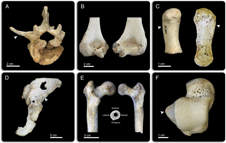
Current knowledge of the evolution of the postcranial skeleton in the genus Homo is hampered by a geographically and chronologically scattered fossil record. Here we present a complete characterization of the postcranium of the middle Pleistocene paleodeme from the Sima de los Huesos (SH) and its paleobiological implications. The SH hominins show the following: (i) wide bodies, a plesiomorphic character in the genus Homo inherited from their early hominin ancestors; (ii) statures that can be found in modern human middle-latitude populations that first appeared 1.6-1.5 Mya; and (iii) large femoral heads in some individuals, a trait that first appeared during the middle Pleistocene in Africa and Europe. The intrapopulational size variation in SH shows that the level of dimorphism was similar to modern humans (MH), but the SH hominins were less encephalized than Neandertals. SH shares many postcranial anatomical features with Neandertals. Although most of these features appear to be either plesiomorphic retentions or are of uncertain phylogenetic polarity, a few represent Neandertal apomorphies. Nevertheless, the full suite of Neandertal-derived features is not yet present in the SH population. The postcranial evidence is consistent with the hypothesis based on the cranial morphology that the SH hominins are a sister group to the later Neandertals. Comparison of the SH postcranial skeleton to other hominins suggests that the evolution of the postcranium occurred in a mosaic mode, both at a general and at a detailed level.
Keywords: Sierra de Atapuerca; bauplan; human evolution; phylogeny; postcranial anatomy.
Conflict of interest statement
The authors declare no conflict of interest.
Figures

References
-
- Wood B, Collard M. The human genus. Science. 1999;284(5411):65–71. - PubMed
-
- Abitbol MM. Lateral view of Australopithecus afarensis: Primitive aspects of bipedal positional behavior in the earliest hominids. J Hum Evol. 1995;28(3):211–229.
-
- Ruff C. Relative limb strength and locomotion in Homo habilis. Am J Phys Anthropol. 2009;138(1):90–100. - PubMed
-
- Haeusler M, McHenry HM. Body proportions of Homo habilis reviewed. J Hum Evol. 2004;46(4):433–465. - PubMed
Publication types
MeSH terms
LinkOut - more resources
Full Text Sources
Other Literature Sources
