. 2015 Sep 15;112(37):E5142-9.
doi: 10.1073/pnas.1507726112. Epub 2015 Aug 31.

Balancing between affinity and speed in target DNA search by zinc-finger proteins via modulation of dynamic conformational ensemble

Affiliations

Balancing between affinity and speed in target DNA search by zinc-finger proteins via modulation of dynamic conformational ensemble

Levani Zandarashvili et al. Proc Natl Acad Sci U S A. .

Abstract

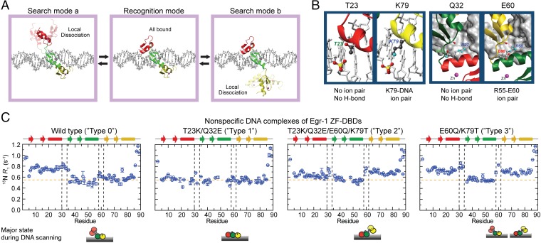
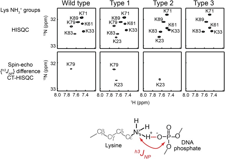
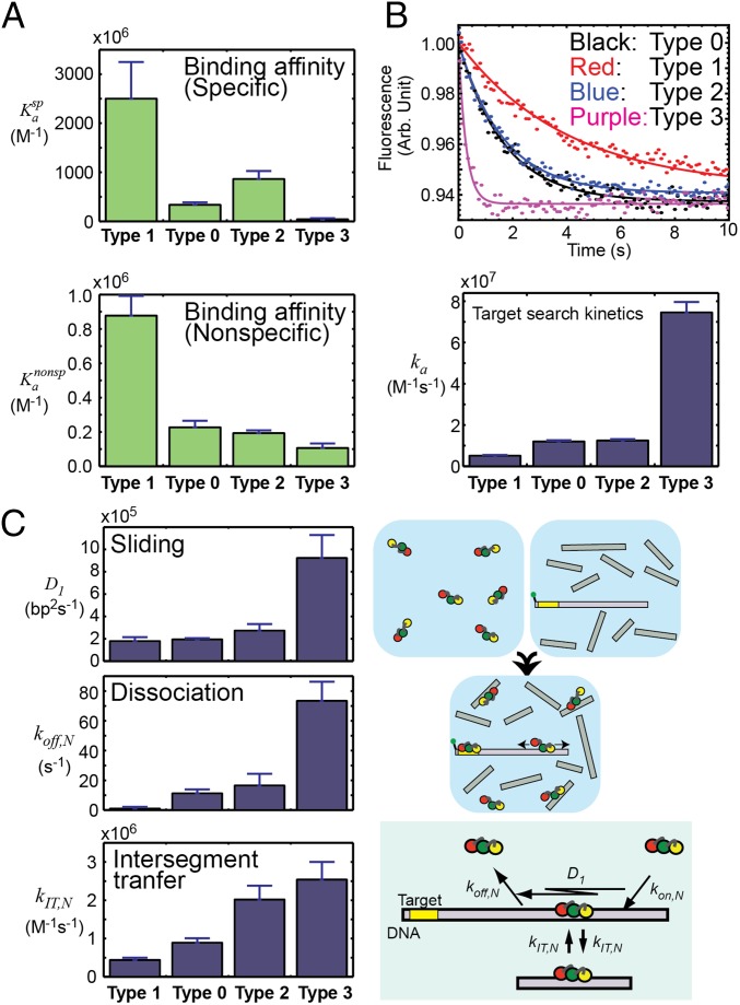
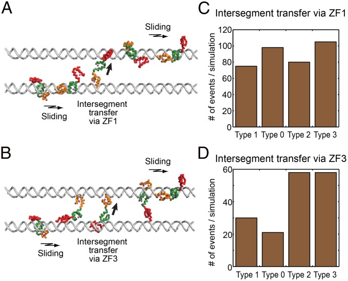
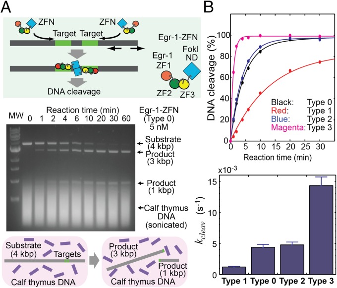
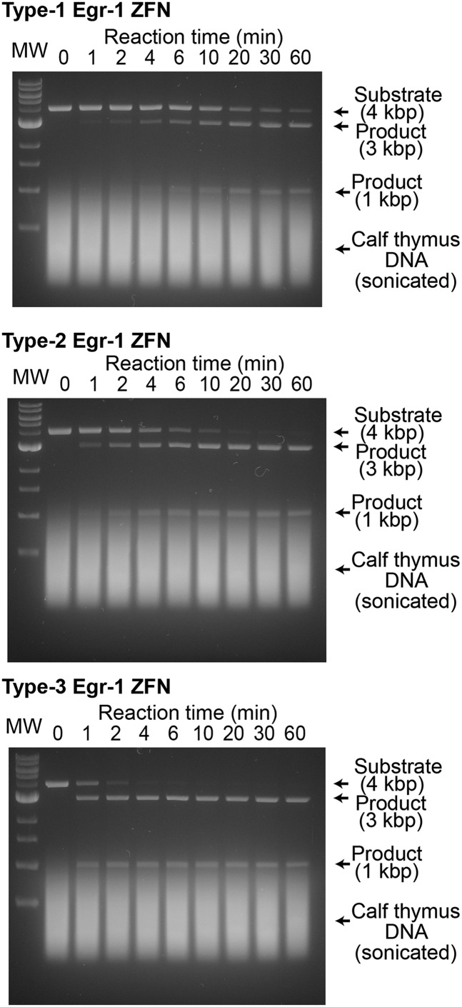
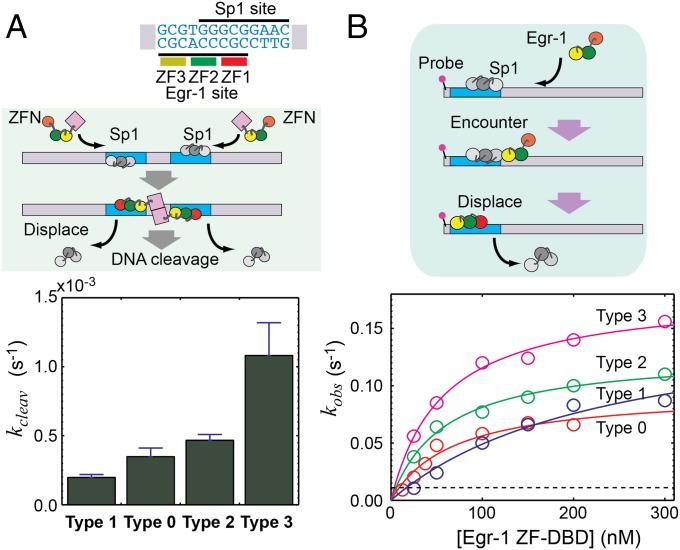
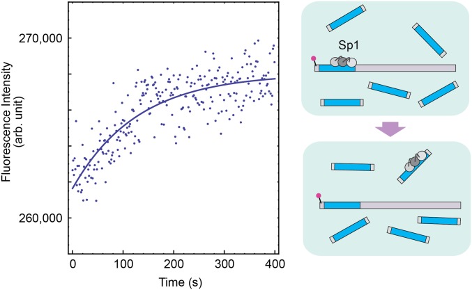
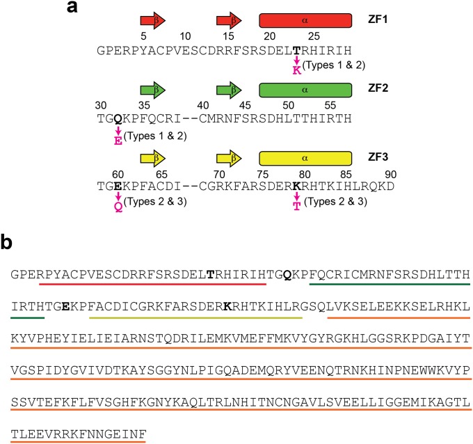
Although engineering of transcription factors and DNA-modifying enzymes has drawn substantial attention for artificial gene regulation and genome editing, most efforts focus on affinity and specificity of the DNA-binding proteins, typically overlooking the kinetic properties of these proteins. However, a simplistic pursuit of high affinity can lead to kinetically deficient proteins that spend too much time at nonspecific sites before reaching their targets on DNA. We demonstrate that structural dynamic knowledge of the DNA-scanning process allows for kinetically and thermodynamically balanced engineering of DNA-binding proteins. Our current study of the zinc-finger protein Egr-1 (also known as Zif268) and its nuclease derivatives reveals kinetic and thermodynamic roles of the dynamic conformational equilibrium between two modes during the DNA-scanning process: one mode suitable for search and the other for recognition. By mutagenesis, we were able to shift this equilibrium, as confirmed by NMR spectroscopy. Using fluorescence and biochemical assays as well as computational simulations, we analyzed how the shifts of the conformational equilibrium influence binding affinity, target search kinetics, and efficiency in displacing other proteins from the target sites. A shift toward the recognition mode caused an increase in affinity for DNA and a decrease in search efficiency. In contrast, a shift toward the search mode caused a decrease in affinity and an increase in search efficiency. This accelerated site-specific DNA cleavage by the zinc-finger nuclease, without enhancing off-target cleavage. Our study shows that appropriate modulation of the dynamic conformational ensemble can greatly improve zinc-finger technology, which has used Egr-1 (Zif268) as a major scaffold for engineering.

Keywords: DNA scanning; dynamics; kinetics; protein–DNA interactions; target search.


Conflict of interest statement

The authors declare no conflict of interest.

Figures

Fig. 1.
Fig. 1.
Modulation of the search and recognition modes during DNA scanning by the Egr-1 ZF-DBD. (A) Search and recognition modes. (B) Mutation sites used to modulate the balance between the search and recognition modes. (C) Backbone amide 15N R1 relaxation rates measured at the 1H frequency of 800 MHz for the nonspecific complexes of the wild-type and mutant proteins of Egr-1 ZF-DBD with 28-bp DNA duplex, dGTACCGATTGCAGATTCCGAACCTTCAG, which contains neither specific nor semispecific sequences.
Fig. S1.
Fig. S1.
Engineering of intermolecular ion pairs between ZF and DNA as analyzed by 15N-31P scalar coupling h3JNP across a hydrogen bond. h3JNP data for the lysine side-chain NH3+ groups in the specific DNA complex of the Egr-1 ZF-DBDs were analyzed. The 1H-15N HISQC (63) and spin-echo h3JNP modulation difference constant-time HISQC (36) spectra for lysine NH3+ groups of each Egr-1 ZF-DBD bound to 12-bp target DNA are shown. The NH3+ resonances of this complex were assigned as previously (64). The spin-echo h3JNP modulation difference constant-time HISQC spectra show signals only for lysine NH3+ groups that form a hydrogen bond with DNA phosphate and exhibit sizable 15N-31P coupling across the hydrogen bond of the intermolecular ion pair (36). Because of the K79T mutation, the type 2 and type 3 mutant complexes do not show signals from K79. Because of the T23K mutation, type 1 and type 2 mutant complexes do show additional signals from K23. The presence of the h3JNP coupling for the K23 NH3+ group represents direct evidence of the intermolecular ion pair artificially introduced for the type 1 and type 2 mutant complexes (Fig. 1B).
Fig. S2.
Fig. S2.
15N longitudinal relaxation rates R1 measured at the 1H frequency of 800 MHz for protein backbone amide groups of the specific complexes of Egr-1 ZF-DBDs with 28-bp DNA containing a target sequence. In contrast to the data for the nonspecific complexes shown in Fig. 1C, these specific complexes exhibit virtually the same 15N R1 profiles for the four constructs of Egr-1 ZF-DBDs.
Fig. S3.
Fig. S3.
Impact of the T23K, Q32E, E60Q, and K79T mutations on the interdomain dynamics of the free Egr-1 ZF-DBD. (A) 15N longitudinal relaxation rates R1 measured for protein backbone amide groups of the Egr-1 ZF-DBDs. For all four constructs of types 0–3, ZF1 and ZF3 exhibited larger R1 rates than ZF2, presumably because of less restricted domain motions (i.e., one linkage vs. two linkages). It should be noted that the 15N R1 profiles are similar for all of the four proteins in the free state. (B) SAXS data for the type 1 (red) and type 3 (green) mutant proteins in the free state. The SAXS data were collected with a Rigaku BioSAXS-1000 instrument using 4 mg/mL protein in the same buffer as that used for the NMR experiments. Intensity I is plotted as a function of the scattering vector q with logarithmic scales for both axes. The SAXS data for these proteins were statistically identical. These NMR and SAXS data indicate that the mutations do not significantly impact the interdomain dynamics of the Egr-1 ZF-DBD in the free state.
Fig. 2.
Fig. 2.
Trade-off between target search efficiency and binding affinity of Egr-1 ZF-DBD. Data for the four constructs of Egr-1 ZF-DBD are compared. (A) Affinities for specific and nonspecific 12-bp DNA duplexes at 150 mM KCl. Association constants in M−1 are shown. Also see Table S1. (B) Target search kinetics analyzed by the stopped-flow fluorescence assay in which the protein was mixed at 150 mM KCl with a solution of fluorescence-labeled DNA (2.5 nM) and sonicated calf thymus DNA (56 μM base pairs) as competitors in large excess. (C) One-dimensional diffusion coefficient D1 for sliding, the dissociation rate constant koff,N for dissociation from nonspecific DNA, and the kinetic rate constant kIT,N for intersegment transfer between two nonspecific sites on district DNA duplexes. These kinetic parameters for translocation of the protein on DNA were measured as previously described (26, 27).
Fig. 3.
Fig. 3.
Intersegment transfer observed in coarse-grained dynamics simulations for the four types of Egr-1 ZF-DBD constructs. (A) Snapshots for type 0 Egr-1 ZF-DBD. Intersegment transfer takes place mostly via ZF1. (B) Snapshots for type 3 Egr-1 ZF-DBD. Intersegment transfer takes place via both ZF1 and ZF3. (C) Frequency of intersegment transfer via ZF1. (D) Frequency of intersegment transfer via ZF3. The number of intersegmental transfer events was probed at salt concentration of 75 mM.
Fig. 4.
Fig. 4.
Enhancement of sequence-specific DNA-cleavage efficiency of Egr-1 ZFN by improving the target search efficiency. (A) Site-specific DNA cleavage by Egr-1 ZFN (type 0). The corresponding data for the other constructs are shown in Fig. S4. Reaction mixtures quenched at different times were subjected to 0.9% agarose/TBE gel electrophoresis. (B) Time courses of the site-specific DNA cleavage and the DNA-cleavage rates for the type 0, 1, 2, and 3 Egr-1 ZFNs.
Fig. S4.
Fig. S4.
Sequence-specific DNA cleavage by type 1, 2, and 3 Egr-1 ZFNs. The corresponding data for type 0 Egr-1 ZFN is shown in Fig. 4A. Each assay of site-specific DNA cleavage by Egr-1 ZFN (5 nM) was carried out at 22 °C for a 900-μL solution of this linear 4-kbp substrate DNA (1 nM) in a buffer of 20 mM Tris⋅HCl (pH 7.5), 150 mM KCl, 0.1 mM ZnCl2, 1 mM MgCl2, 2 mM β-mercaptoethanol, 5% glycerol, and 37 μg/mL competitor DNA (corresponding to 56 μM base pairs). Sonicated calf thymus DNA (average length, ∼0.5 kbp) was used as the competitor. A 100-μL aliquot of the reaction mixture was sampled and quenched at distinct time points (0, 1, 2, 4, 6, 10, 20, 30, and 60 min). The lanes marked “MW” are for the molecular weight makers (NEB 1-kb DNA ladder) including 0.5, 1.0, 1.5, 2.0, 3.0, 4.0, 5.0, 6.0, 8.0, and 10.0-kbp DNA. Results of the kinetic analysis are shown in Fig. 4B.
Fig. S5.
Fig. S5.
Off-target DNA cleavage by type 0, 1, 2, and 3 Egr-1 ZFNs at a high concentration. This experiment was performed using the same experimental conditions as those used for Fig. 4 and Fig. S4, except that 50 nM Egr-1 ZFN was used instead of 5 nM. The reaction time was 60 min for each. DNA bands indicated by red arrows correspond to the products of the off-target DNA cleavage.
Fig. 5.
Fig. 5.
Displacement of the Sp1 ZF-DBD from the overlapping target sites. (A) Kinetics of the site-specific DNA cleavage by Egr-1 ZFNs in the presence the Sp1 ZF-DBD bound to the overlapping target sites. This assay was conducted using the Egr-1 ZFN (5 nM), 28-bp nonspecific competitor DNA (2,000 nM), and 4-kbp substrate DNA (1 nM) containing Egr-1/Sp1 overlapping sites, which Sp1 ZF-DBD (200 nM) initially occupied. (B) Kinetics of the Egr-1 ZF-DBDs' target association that requires displacement of Sp1 ZF-DBD from the overlapping target site. Apparent pseudo-first-order rate constants are plotted as a function of the concentrations of Egr-1 ZF-DBDs. Solid lines represent the best-fit curves obtained by hyperbolic fitting (see Materials and Methods). A dotted line represents the value of the rate constant for Sp1’s spontaneous dissociation from the overlapping site in the absence of Egr-1 ZF-DBDs (Fig. S6).
Fig S6.
Fig S6.
Kinetics of dissociation of Sp1 ZF-DBD from the FAM-labeled 117-bp DNA in the absence of Egr-1 ZF-DBD. In this experiment, the FAM fluorescence was monitored immediately after a solution of the FAM-labeled 117-bp DNA (5 nM) and Sp1 ZF-DBD (50 nM) was mixed with a solution of unlabeled 28-bp DNA containing the overlapping target sites (2,000 nM). Before mixing these two solutions, the target site of the FAM-labeled 117-bp DNA was initially bound to Sp1 ZF-DBD. Once dissociated from the FAM-labeled DNA, Sp1 ZF-DBD binds to the unlabeled 28-bp DNA. This transfer via dissociation increases the FAM fluorescence intensity. In fact, this increase was of the same magnitude as the fluorescence quenching observed upon Sp1’s binding to the probe DNA. Compared with the change upon binding of Egr-1 ZF-DBD to the Egr-1 target site on the same DNA, the change in fluorescence intensity upon binding of Sp1 is relatively small because the Sp1 site is apart by 5 bp from the 5′ terminus to which FAM is attached. The buffer conditions were the same as those used for Fig. 5B. By monoexponential fitting for the data of 9 replicates, the rate constant koff,Sp1 for spontaneous dissociation of Sp1 from the overlapping Egr-1/Sp1 target site was determined to be 0.011 ± 0.001 s−1.
Fig S7.
Fig S7.
The Egr-1 ZF protein constructs used in this study. (A) The amino acid sequence of the Egr-1 ZF-DBD constructs. Each construct is a 91-residue protein containing 3 ZFs. The first two residues are from the expression vector. The residue numbering is according to Pavletich and Pabo (37). The sequences of individual ZFs are aligned, and the mutation sites for type 1, 2, and 3 mutant proteins are indicated. (B) The amino acid sequence of the type 0 Egr-1 ZFN. Regions of Egr-1 ZF1, ZF2, ZF3, and FokI ND are indicated by red, green, yellow, and orange lines, respectively. The mutation sites for type 1, 2, and 3 constructs are shown in bold.

Similar articles

Cited by

References

    1. Choo Y, Isalan M. Advances in zinc finger engineering. Curr Opin Struct Biol. 2000;10(4):411–416. - PubMed
    1. Pabo CO, Peisach E, Grant RA. Design and selection of novel Cys2His2 zinc finger proteins. Annu Rev Biochem. 2001;70:313–340. - PubMed
    1. Segal DJ, Barbas CF., 3rd Custom DNA-binding proteins come of age: Polydactyl zinc-finger proteins. Curr Opin Biotechnol. 2001;12(6):632–637. - PubMed
    1. Durai S, et al. Zinc finger nucleases: Custom-designed molecular scissors for genome engineering of plant and mammalian cells. Nucleic Acids Res. 2005;33(18):5978–5990. - PMC - PubMed
    1. Urnov FD, Rebar EJ, Holmes MC, Zhang HS, Gregory PD. Genome editing with engineered zinc finger nucleases. Nat Rev Genet. 2010;11(9):636–646. - PubMed

Publication types