. 2015 Sep 1;11(9):e1004398.
doi: 10.1371/journal.pcbi.1004398. eCollection 2015 Sep.

An Integrated Framework Advancing Membrane Protein Modeling and Design

Affiliations

An Integrated Framework Advancing Membrane Protein Modeling and Design

Rebecca F Alford et al. PLoS Comput Biol. .

Abstract

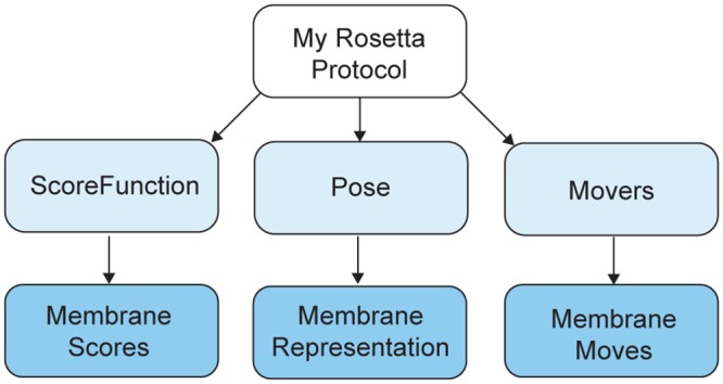
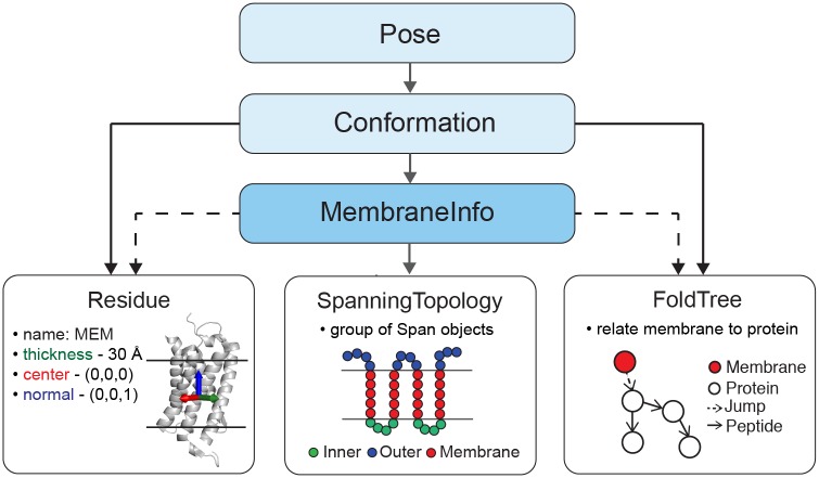
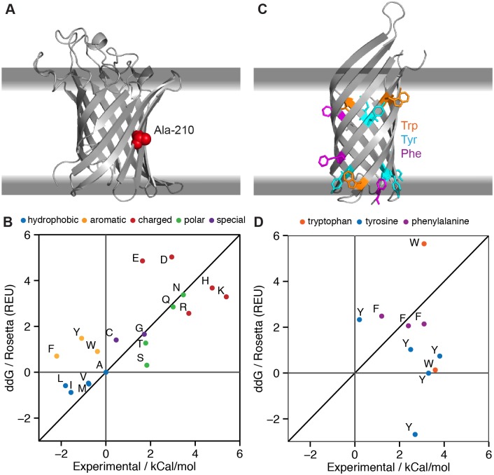
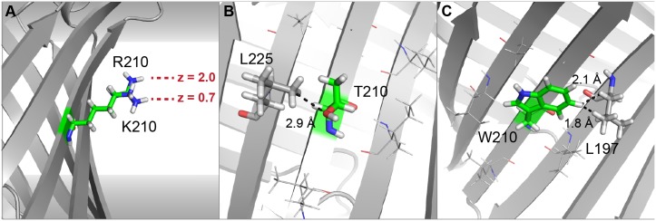
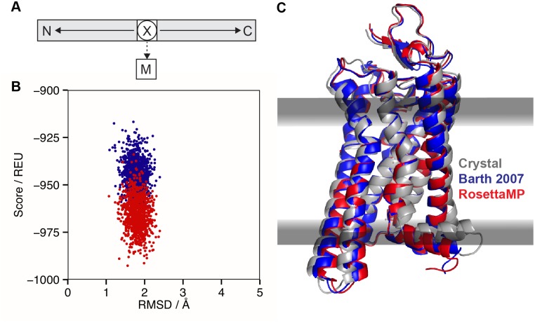
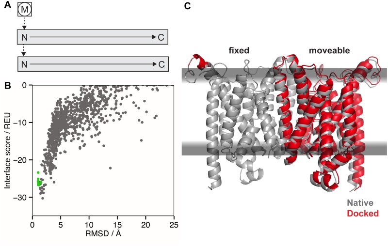
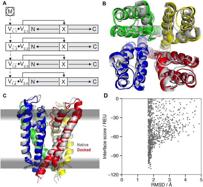
Membrane proteins are critical functional molecules in the human body, constituting more than 30% of open reading frames in the human genome. Unfortunately, a myriad of difficulties in overexpression and reconstitution into membrane mimetics severely limit our ability to determine their structures. Computational tools are therefore instrumental to membrane protein structure prediction, consequently increasing our understanding of membrane protein function and their role in disease. Here, we describe a general framework facilitating membrane protein modeling and design that combines the scientific principles for membrane protein modeling with the flexible software architecture of Rosetta3. This new framework, called RosettaMP, provides a general membrane representation that interfaces with scoring, conformational sampling, and mutation routines that can be easily combined to create new protocols. To demonstrate the capabilities of this implementation, we developed four proof-of-concept applications for (1) prediction of free energy changes upon mutation; (2) high-resolution structural refinement; (3) protein-protein docking; and (4) assembly of symmetric protein complexes, all in the membrane environment. Preliminary data show that these algorithms can produce meaningful scores and structures. The data also suggest needed improvements to both sampling routines and score functions. Importantly, the applications collectively demonstrate the potential of combining the flexible nature of RosettaMP with the power of Rosetta algorithms to facilitate membrane protein modeling and design.


Conflict of interest statement

The authors have declared that no competing interests exist.

Figures

Fig 1
Fig 1. RosettaMP directly extends the architecture of Rosetta3.
Every Rosetta protocol requires at least these three main objects for modeling or design tasks (light blue): a Pose to a represent a biomolecule, a ScoreFunction to rank modeled structures and sequences, and Movers to sample new conformations of the Pose. RosettaMP directly extends this architecture (blue) by adding an element to the Pose representing the membrane bilayer, restructuring the original membrane ScoreFunction to rely on this membrane representation, and implementing a new set of Movers to sample the conformational search space available in the membrane bilayer.
Fig 2
Fig 2. Detailed architecture of RosettaMP.
RosettaMP represents the membrane bilayer using three main components connected to a central MembraneInfo object (blue). MembraneInfo stores information needed to represent the membrane (line arrows) and tracks information present in the Pose (dotted arrows). A special Residue type is added to the Pose, describing the geometry of the membrane bilayer by coordinates storing the center, normal and thickness of the bilayer. A SpanningTopology object describes the transmembrane regions of the Pose. The FoldTree uses a jump edge to establish the connection between the membrane residue and the protein. MembraneInfo is also a central repository for membrane-related features such as lipid accessibility of each residue (LipidAccInfo). A full Universal Markup Language (UML) diagram is presented in Fig A in S1 File.
Fig 3
Fig 3. PyRosetta script for calculating the ΔΔG of mutation via RosettaMP.
This example script loads Rosetta, adds the membrane representation, and uses the membrane score function to compute the ΔΔG of mutation in the membrane. Left: Python script used for ΔΔG calculations. Right: calc_ddG method used for computing ΔΔG of mutation.
Fig 4
Fig 4. MPddG computes free energy changes upon mutation in the membrane environment (ΔΔG).
(A) Outer membrane protein phospholipase A (OmpLA, PDB 1qd6) with its native alanine at position 210 in red at the center of the membrane. (B) Plot of RosettaMP-calculated fixed-backbone ΔΔGs versus experimentally measured values of Moon & Fleming for variants at position 210 [50]. Proline is off-scale (ΔΔG pred = 193.2 REU) due to incompatible backbone torsions yielding ring closure penalties. (C) Outer membrane protein A (OmpA, PDB 1qjp) with aromatic residues mutated to alanine at various interfacial positions. (D) Plot of RosettaMP-calculated ΔΔGs versus experimentally measured values of Hong & Tamm [51]. The mutation W15A is off-scale (ΔΔG pred = -43.0 REU) due to the loss of repulsive interactions upon mutation to alanine. Both (B) and (D) include a line for y = x.
Fig 5
Fig 5. Structures of mutant residues at position 210 of OmpLA.
(A) The charged residues arginine and lysine (superimposed) cannot reach the interface region. The z-coordinate shows the difference in membrane depth of the two charged side chains. Membrane environment scores are unfavorable for both, with lysine being slightly more unfavorable. (B) Insertion of threonine at position 210 is penalized by a mild clash from the neighboring leucine 225; serine at this position is accommodated more easily (Fig D in S1 File). (C) The tryptophan side chain is close to the neighboring leucine 197, resulting in large repulsive scores. All aromatic mutations have a comparably large repulsive van der Waals and rotamer scores, resulting in over-prediction of their ΔΔG values.
Fig 6
Fig 6. MPrelax for high-resolution refinement of a membrane protein.
(A) FoldTree representation for the MPrelax protocol with the residue closest to the center-of-mass of the protein being at the root of the FoldTree (circled X). The membrane residue (M) is attached via a flexible jump edge (dashed arrow). Protein chains are shown as gray boxes with N- and C- termini marked and peptide edges shown as solid arrows. (B) Rosetta total score vs. backbone RMSD to the crystal structure for 1000 models of meta-rhodopsin. Models in blue are created with the original membrane relax protocol of RosettaMembrane; models in red are created with MPrelax. (C) Crystal structure of meta-rhodopsin in gray (PDB 3pxo) superimposed with the lowest scoring models from both the original RosettaMembrane protocol (blue) and the MPrelax protocol (red).
Fig 7
Fig 7. Protein-protein docking in the membrane bilayer using MPdock.
(A) FoldTree representation used in MPdock with the membrane residue (M) being fixed at the root (circled) of the FoldTree and the protein chains as docking partners attached via jump edges. (B) Interface score vs. backbone RMSD to the native structure for 1000 models of the vitamin B12 importer BtuCD. The RMSD is the ‘ligand’ RMSD, which is computed only over the moving partner after superimposing the fixed partner and membrane. The green dots represent ten models created by minimizing the crystal structure. The interface score of the crystal structure (180.5 REU) is outside of the plotting range due to clashes. (C) Native structure of the vitamin B12 importer (gray, PDB 2qi9) superimposed with the model having the lowest interface score (red).
Fig 8
Fig 8. Assembly of symmetric protein complexes in the membrane using MPsymdock.
(A) FoldTree representation of the homo-tetrameric KcsA potassium channel with the membrane residue (M) at the root (circled). The virtual residues V1,i and V2,i required for the symmetry machinery are described in the text. (B) Native structure in gray (PDB 1bl8) superimposed with the model from MPsymdock with the lowest interface score. The view is from the extracellular side of the membrane. (C) Membrane plane view of (B). (D) Interface score vs. backbone RMSD to the native structure for 1000 models of the KcsA potassium channel. The lowest scoring model, shown in (B) and (C), is indicated in red.

References

    1. Von Heijne G. The membrane protein universe: what’s out there and why bother? J Intern Med. 2007;261: 543–557. 10.1111/j.1365-2796.2007.01792.x - DOI - PubMed
    1. Tan S, Tan HT, Chung MCM. Membrane proteins and membrane proteomics. Proteomics. 2008;8: 3924–3932. 10.1002/pmic.200800597 - DOI - PubMed
    1. Bakheet TM, Doig AJ. Properties and identification of human protein drug targets. Bioinformatics. 2009;25: 451–457. 10.1093/bioinformatics/btp002 - DOI - PubMed
    1. Overington JP, Al-Lazikani B, Hopkins AL. How many drug targets are there? Nat Rev Drug Discov. 2006;5: 993–996. 10.1038/nrd2199 - DOI - PubMed
    1. Berman HM, Westbrook J, Feng Z, Gilliland G, Bhat TN, Weissig H, et al. The Protein Data Bank. Nucleic Acids Res. 2000;28: 235–242. 10.1093/nar/28.1.235 - DOI - PMC - PubMed

Publication types

MeSH terms

Substances