. 2015 Sep 1;10(9):e0136851.
doi: 10.1371/journal.pone.0136851. eCollection 2015.

Ability to Generate Patient-Derived Breast Cancer Xenografts Is Enhanced in Chemoresistant Disease and Predicts Poor Patient Outcomes

Affiliations

Ability to Generate Patient-Derived Breast Cancer Xenografts Is Enhanced in Chemoresistant Disease and Predicts Poor Patient Outcomes

Priscilla F McAuliffe et al. PLoS One. .

Erratum in

Abstract

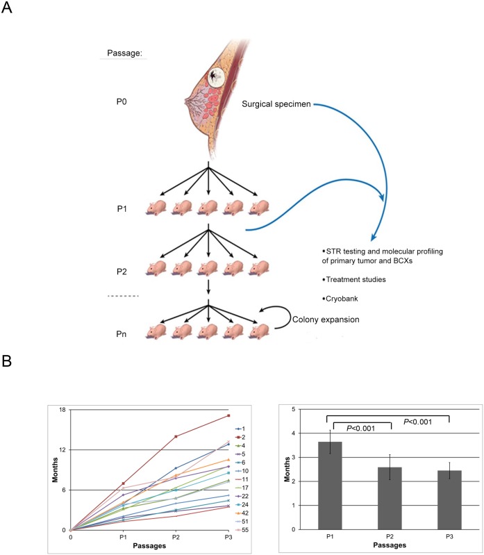
Background: Breast cancer patients who are resistant to neoadjuvant chemotherapy (NeoCT) have a poor prognosis. There is a pressing need to develop in vivo models of chemo resistant tumors to test novel therapeutics. We hypothesized that patient-derived breast cancer xenografts (BCXs) from chemo- naïve and chemotherapy-exposed tumors can provide high fidelity in vivo models for chemoresistant breast cancers.

Methods: Patient tumors and BCXs were characterized with short tandem repeat DNA fingerprinting, reverse phase protein arrays, molecular inversion probe arrays, and next generation sequencing.

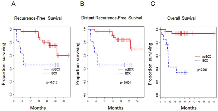
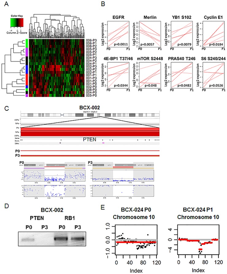
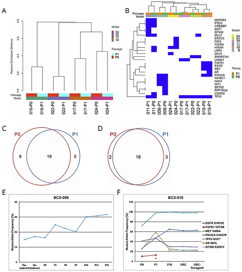
Results: Forty-eight breast cancers (24 post-chemotherapy, 24 chemo-naïve) were implanted and 13 BCXs were established (27%). BCX engraftment was higher in TNBC compared to hormone-receptor positive cancer (53.8% vs. 15.6%, p = 0.02), in tumors from patients who received NeoCT (41.7% vs. 8.3%, p = 0.02), and in patients who had progressive disease on NeoCT (85.7% vs. 29.4%, p = 0.02). Twelve patients developed metastases after surgery; in five, BCXs developed before distant relapse. Patients whose tumors developed BCXs had a lower recurrence-free survival (p = 0.015) and overall survival (p<0.001). Genomic losses and gains could be detected in the BCX, and three models demonstrated a transformation to induce mouse tumors. However, overall, somatic mutation profiles including potential drivers were maintained upon implantation and serial passaging. One BCX model was cultured in vitro and re-implanted, maintaining its genomic profile.

Conclusions: BCXs can be established from clinically aggressive breast cancers, especially in TNBC patients with poor response to NeoCT. Future studies will determine the potential of in vivo models for identification of genotype-phenotype correlations and individualization of treatment.


Conflict of interest statement

Competing Interests: The authors have declared that no competing interests exist.

Figures

Fig 1
Fig 1. Generating and maintaining patient-derived breast cancer xenografts (BCXs) and their time to passage.
(A) After surgery, patient tumors (P0) were implanted into nude mice, creating a patient-derived BCX, passage 1 (P1). When tumors reached 1.5 cm diameter, they were harvested and implanted into 5 new mice (P2), and subsequent passages respectively. Patient tumors and BCXs were evaluated by STR and selected passages underwent molecular and histologic characterization. (B) Y-axis depicts time to reach 1.5 cm with each passage. The graph shows thirteen BCXs that were serially passage. (C) Time to passage (in months) at from implantation to first passage (P1), P1 to P2 (P2), and P1 to P3 (P3). Time to passage at P2 and P3 were compared to time to passage at from implantation to first passage (P1).
Fig 2
Fig 2. Survival Outcomes in Patients Based on BCX development.
(A) Recurrence-free survival (in months) in patients whose tumors developed BCX versus those did not (no BCX). (B) Distant recurrence-free survival (in months) in patients whose tumors developed BCX versus those did not (no BCX). (C) Overall survival (in months) in patients whose tumors developed BCX versus those did not (no BCX).
Fig 3
Fig 3. Molecular differences between patients’ tumors and BCXs.
(A) Unsupervised clustering of proteomic profile of patient tumors and BCXs as determined by RPPA. Each protein tested represents a column: Red, high expression; green, low expression. Samples are listed on the right side. Left, cluster trees of sample groups; top, cluster trees of proteins. Each BCX model’s P1-3 generation clustered together, demonstrating relative stability of the proteomic profile once growth in mouse is established. However, all P0 generations clustered together, suggesting that differences between patient tumor-xenograft proteomic profiles is greater than inter-tumoral differences. (B) Selected proteins and phosphoproteins that are differentially expressed between patient tumors (P0) and the first-generation of BCXs passaged through nude mice (P1). Protein levels were compared between the two groups with RPPA; all shown have a FDR 0.1 or less. (C) Copy number analysis determined PTEN loss in MDA-BCX-002. Top panel shows the chromosome 10 ideogram and the PTEN gene. Deletions are plotted in red below the 0% baseline, and dark red indicates homozygous loss. The lowest portion of the top panel separates out P0 and P3. A heterozygous PTEN loss is detected in P0, and in the P3, the second PTEN allele is lost, resulting in a homozygous PTEN loss. Bottom panel shows the PTEN gene ideogram followed by the copy number aberration plot and the allele frequency plot for P0 and P3. A heterozygous PTEN loss is detected in P0 (single red line), and a homozygous PTEN loss is detected in P3 (double red line). Each blue dot corresponds to an individual probe on the array. The brown and purple lines mark the thresholds for loss of heterozygosity (LOH) and allelic imbalance regions, respectively. (D) PCR confirmed PTEN deletion in genomic DNA from Patient 2 tumor and BCX-002 P3. PTEN was undetectable in P3 but present in P0. RB1, another tumor suppressor gene, is detected in both samples and included for comparison. (E) PTEN loss demonstrated by next generation sequencing in BCX-024. P0 on the left, P1 on the right.
Fig 4
Fig 4. Analysis of mutation data.
(A) Clustering of whole exome sequencing mutation data based on the mutation status of the genes in P0 and P1 samples of four models. (B) Clustering of targeted exome sequencing data based on the mutation status of 201 genes in P0 and P1 samples of six models. (C) Venn diagram of mutations in P0 and P1 samples of six models on the targeted exome sequencing platform. (D) Venn diagram of high allelic frequency mutations (10% or higher) on targeted exome sequencing in P0 and P1 samples of six models. Allele frequency cutoff was 10%. (E) Genomic stability of PIK3CA mutation in BCX-006 model. Fine needle aspiration biopsy samples of before and after 12 weeks of neoadjuvant chemotherapy (including a rapalog) were sequenced by ion torrent. Patient’s P0 tumor and BCX-006 P1 tumor and subsequent passages were analyzed by targeted exome sequencing. PIK3CA H1047R allele frequencies are presented. (F) Conditionally reprogrammed cells (CRC) derived from BCX-010 were passaged four times in vitro and then injected into mice flanks. The cultured cells and CRC-derived xenografts maintained a mutation profile similar to the originating PDX.

References

    1. Caudle AS, Gonzalez-Angulo AM, Hunt KK, Liu P, Pusztai L, Symmans WF, et al. Predictors of tumor progression during neoadjuvant chemotherapy in breast cancer. J Clin Oncol. 2010;28(11):1821–8. Epub 2010/03/17. doi: JCO.2009.25.3286 [pii] 10.1200/JCO.2009.25.3286 - DOI - PMC - PubMed
    1. Kuerer HM, Newman LA, Smith TL, Ames FC, Hunt KK, Dhingra K, et al. Clinical course of breast cancer patients with complete pathologic primary tumor and axillary lymph node response to doxorubicin-based neoadjuvant chemotherapy. J Clin Oncol. 1999;17(2):460–9. Epub 1999/03/18. . - PubMed
    1. Symmans WF, Peintinger F, Hatzis C, Rajan R, Kuerer H, Valero V, et al. Measurement of residual breast cancer burden to predict survival after neoadjuvant chemotherapy. J Clin Oncol. 2007;25(28):4414–22. Epub 2007/09/06. doi: JCO.2007.10.6823 [pii] 10.1200/JCO.2007.10.6823 . - DOI - PubMed
    1. Liedtke C, Mazouni C, Hess KR, Andre F, Tordai A, Mejia JA, et al. Response to neoadjuvant therapy and long-term survival in patients with triple-negative breast cancer. J Clin Oncol. 2008;26(8):1275–81. 10.1200/JCO.2007.14.4147 . - DOI - PubMed
    1. Vargo-Gogola T, Rosen JM. Modelling breast cancer: one size does not fit all. Nature reviews. 2007;7(9):659–72. Epub 2007/08/28. doi: nrc2193 [pii] 10.1038/nrc2193 . - DOI - PubMed

Publication types

MeSH terms