Review
. 2015 Dec 10:219:119-128.
doi: 10.1016/j.jconrel.2015.08.049. Epub 2015 Aug 29.

Controlled drug release for tissue engineering

Affiliations
Review

Controlled drug release for tissue engineering

Kunal J Rambhia et al. J Control Release. .

Abstract

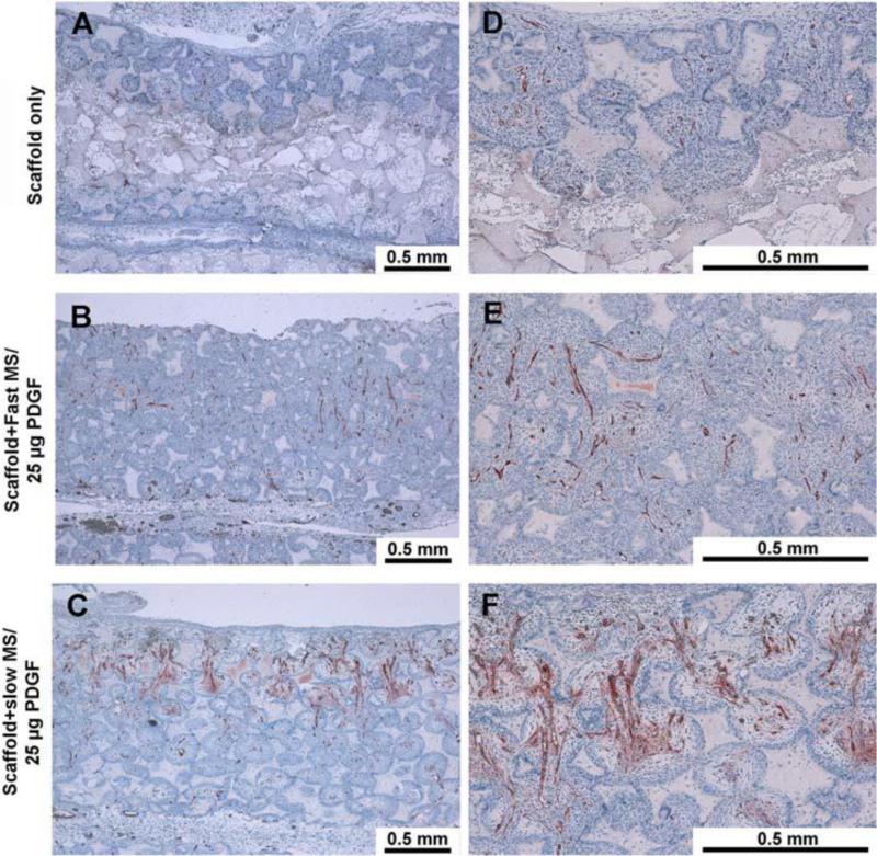
Tissue engineering is often referred to as a three-pronged discipline, with each prong corresponding to 1) a 3D material matrix (scaffold), 2) drugs that act on molecular signaling, and 3) regenerative living cells. Herein we focus on reviewing advances in controlled release of drugs from tissue engineering platforms. This review addresses advances in hydrogels and porous scaffolds that are synthesized from natural materials and synthetic polymers for the purposes of controlled release in tissue engineering. We pay special attention to efforts to reduce the burst release effect and to provide sustained and long-term release. Finally, novel approaches to controlled release are described, including devices that allow for pulsatile and sequential delivery. In addition to recent advances, limitations of current approaches and areas of further research are discussed.

Keywords: Biomaterials; Controlled release; Drug delivery; Polymer; Regenerative medicine; Scaffold; Tissue engineering.


Figures

Figure 1
Figure 1
SEM images of Solid Walled (SW)-PLLA and NF-PLLA scaffolds. (A) Macroporous structure of SW-PLLA scaffolds at low magnification; (B) Solid-walled architecture of SW-PLLA scaffolds at high magnification; (C) Macroporous structure of NF-PLLA scaffolds at low magnification; and (D) Nanofibrous architecture of NF-PLLA scaffolds at high magnification. Reprinted from Biomaterials, 32(31), Jing Wang, Haiyun Ma, Xiaobing Jin, et al. The effect of scaffold architecture on odontogenic differentiation of human dental pulp stem cells. 7822–7830. (2011) with permission from Elsevier.
Figure 2
Figure 2
Nanofibrous scaffolds with PDGF microspheres promote vasculogenesis in vivo. Left panel is low magnification (10X), right panel is high magnification (40X). Positive Factor VIII stained blood vessels were located in the central regions of the pores within penetrated tissues. The blood vessels also permeated through the inter-openings between each pore. The group with 25 mg PDGF encapsulated in slow release PLGA microspheres had measurably more vascularization. Reprinted from PLoS One, 3(3), Qiming Jin, Guabao Wei, Zhao Lin, et. al. Nanofibrous Scaffolds Incorporating PDGF-BB Microspheres Induce Chemokine Expression and Tissue Neogenesis In Vivo. e1729 (2008), under Creative Commons Attribution license
Figure 3
Figure 3
Characterization of PLGA50–64K nanospheres (NS) and nanosphere incorporated PLLA nano-fibrous scaffolds (NS-scaffolds). (A) Scanning electron micrograph of rhBMP-7 containing PLGA50–64K nanospheres; (B) Macroscopic photographs of PLLA scaffolds before (left) and after (right) nanosphere incorporation; (C, D) Scanning electron micrographs of PLLA nano-fibrous scaffolds before nanosphere incorporation at 100× (C) and 10,000× (D); (E, F) Scanning electron micrographs of PLLA nano-fibrous scaffolds after PLGA50–64K nanosphere incorporation at 100× (E) and 10,000× (F). Reprinted from Biomaterials, 28 (12), Guobao Wei, Qiming Jin, William V. Giannobile, Peter X. Ma The enhancement of osteogenesis by nano-fibrous scaffolds incorporating rhBMP-7 nanospheres. 2087-2096. (2007) with permission from Elsevier.

References

    1. Motamedian SR, Hosseinpour S, Ahsaie MG, Khojasteh A. Smart scaffolds in bone tissue engineering : A systematic review of literature. World J Stem Cells. 2015;7(3):657–668. doi:10.4252/wjsc.v7.i3.657. - PMC - PubMed
    1. Hu J, Ma PX. Nano-fibrous tissue engineering scaffolds capable of growth factor delivery. Pharm Res. 2011;28(6):1273–1281. doi:10.1007/s11095-011-0367-z. - PMC - PubMed
    1. Ma G. Microencapsulation of protein drugs for drug delivery: strategy, preparation, and applications. J Control Release. 2014;193:324–340. doi:10.1016/j.jconrel.2014.09.003. - PubMed
    1. Li J, Xu L, Liu H, et al. Biomimetic synthesized nanoporous silica@poly(ethyleneimine)s xerogel as drug carrier: Characteristics and controlled release effect. Int J Pharm. 2014;467:9–18. doi:10.1016/j.ijpharm.2014.03.045. - PubMed
    1. Mulyasasmita W, Cai L, Dewi RE, et al. Avidity-controlled hydrogels for injectable co-delivery of induced pluripotent stem cell-derived endothelial cells and growth factors. J Control Release. 2014;191:71–81. doi:10.1016/j.jconrel.2014.05.015. - PMC - PubMed

Publication types