. 2015 Sep 1;4(9):e248.
doi: 10.1038/mtna.2015.21.

Effective Small Interfering RNA Therapy to Treat CLCN7-dependent Autosomal Dominant Osteopetrosis Type 2

Affiliations

Effective Small Interfering RNA Therapy to Treat CLCN7-dependent Autosomal Dominant Osteopetrosis Type 2

Mattia Capulli et al. Mol Ther Nucleic Acids. .

Abstract

In about 70% of patients affected by autosomal dominant osteopetrosis type 2 (ADO2), osteoclast activity is reduced by heterozygous mutations of the CLCN7 gene, encoding the ClC-7 chloride/hydrogen antiporter. CLCN7(G215R)-, CLCN7(R767W)-, and CLCN7(R286W)-specific siRNAs silenced transfected mutant mRNA/EGFP in HEK293 cells, in RAW264.7 cells and in human osteoclasts, with no change of CLCN7(WT) mRNA and no effect of scrambled siRNA on the mutant transcripts. Osteoclasts from Clcn7(G213R) ADO2 mice showed reduced bone resorption, a condition rescued by Clcn7(G213R)-specific siRNA. Treatment of ADO2 mice with Clcn7(G213R)-specific siRNA induced increase of bone resorption variables and decrease of trabecular bone mass, leading to an overall improvement of the osteopetrotic bone phenotype. Treatment did not induce overt adverse effects and was effective also with siRNAs specific for other mutants. These results demonstrate that a siRNA-based experimental treatment of ADO2 is feasible, and underscore a translational impact for future strategy to cure this therapeutically neglected form of osteopetrosis.


Figures

Figure 1
Figure 1
In vitro and in vivo tests of CLCN7mutant-specific siRNAs. (a) Cartoon depicting the pEGFP-C1 vector used in the study. (b) HEK293 cells stably transfected with the pEGFP-C1 vector carrying the indicated mutations. Expression of the CLCN7 gene was quantified by real-time RT-PCR on RNA extracted from mutant transfectants, against cells transfected with the empty vector, which did not express CLCN7 mRNA (first bar from left). (c–e) HEK293 cells transfected with the indicated vectors, were treated with the CLCN7mutant-specific siRNA listed in Table 2 as the most effective per each mutation. Concentration-dependent regulation of CLCN7 assessed by real-time RT-PCR, normalized with GAPDH. (f) RT-PCR using primer pairs specific for the Clcn7G213R mRNA showing transcript amplification only in heterozygous (Clcn7G213R/WT) and homozygous (Clcn7G213R/G213R) osteoclasts, while in wild-type osteoclasts (Clcn7WT/WT) no transcript was amplified. (g) Direct DNA sequencing of the amplified transcript shown in f for the Clcn7G213R/WT osteoclasts, demonstrating only the mutant sequence. (h) Osteoclasts generated from the bone marrow mononuclear cells of Clcn7WT/WT and Clcn7G213R/WT mice were treated with the indicated concentration of scrambled (SCR) or Clcn7G213R-specific siRNA. Real-time RT-PCR was performed using the primer pairs specific for the mutant transcript validated in (f) and (g). (i) Osteoclasts were generated from the bone marrow mononuclear cells of Clcn7WT/WT and Clcn7G213R/WT mice onto bone slices and treated with the indicated concentration of SCR and Clcn7G213R-specific siRNA. At the end of experiment, cells were removed by sonication and bone resorption evaluated by the pit assay. (j) Three-month-old Clcn7WT/WT mice were injected once i.p. with 4 mg/kg of Clcn7G213R-sticky siRNA jetPEI conjugate and sacrificed at the indicated time point. Sera were collected and evaluated for total RNA concentration by Nanodrop. (k) Ten-day-old Clcn7G21R/WT mice were injected once i.p. with the indicated doses of SCR- or of Clcn7G213R-sticky siRNA jetPEI conjugate. After 48 hours, mice were sacrificed, RNA was extracted from tibias, and evaluated by real-time RT-PCR using the primer pairs specific for the Clcn7G213R mRNA validated in (f) and (g). In b–e, h–k data are the mean ± SD of three independent experiments or three animals/group. b–e,h,I,k: Student's t-test. j: one-way analysis of variance (ANOVA). For c–e, statistics was also performed by one way ANOVA (shown in Supplementary Table S3).
Figure 2
Figure 2
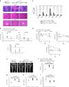
In vivo treatment and safety study. Ten-day-old Clcn7G213R/WT mice were injected i.p. with 4 mg/kg of Clcn7G213R-sticky siRNA jetPEI conjugate, three times a week for 4 weeks. At the end of the experiments, mice were sacrificed and (a) the indicated organs were subjected to histopathological evaluation by hematoxylin/eosin staining (Bar = 100 µm for spleen and kidney, 20 µm for liver). (b) Sera were collected and analyzed by the Reflotron method for the indicated biomarkers of kidney and liver disease, and for the ADO2 biomarker CK. Normal values are between the two dotted lines. (c) RNA was extracted from the indicated organs and subjected to real time RT-PCR using primer pairs specific for the Clcn7G213R mRNA, normalized for gapdh. (d) Ten day-old Clcn7WT/WT and Clcn7G213R/WT were treated with 4 mg/kg of scrambled- (SRC) or Clcn7G213R-sticky siRNA jetPEI conjugate, three times a week for 2 and 4 weeks. At the end of the experiments, mice were sacrificed, then the serum biomarker of bone resorption, CTX, the serum osteoclast biomarker, TRAcP (5b isoform), and the CTX/TRAcP ratio were evaluated after 2 and 4 weeks of treatment. (e) µCT analysis of proximal tibias of mice treated with 4 mg/kg of Clcn7G213R-sticky siRNA jetPEI conjugate, three times a week for 2 weeks, followed by measurements of trabecular (f) bone volume over total tissue volume (BV/TV), (g) trabecular number (Th.N), (h) thickness (Tb.Th), and (i) separation (Tb.Sp). Data are (a,e) representative or (b–d, f–i) the mean ± SD of four to seven mice per group (Student's t-test). For f–i statistics was also performed by one-way analysis of variance (shown in Supplementary Table S3).
Figure 3
Figure 3
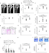
Rescue of the bone phenotype. Ten-day-old Clcn7WT/WT and Clcn7G213R/WT were treated with 4 mg/kg of scrambled- (SRC) or Clcn7G213R-sticky siRNA jetPEI conjugate, three times a week for 4 weeks. At the end of the experiments, mice were sacrificed and their bone phenotype analyzed. (a) µCT analysis of proximal tibias. (b) Trabecular bone volume over total tissue volume (BV/TV). (c) Trabecular number (Tb.N). (d) Trabecular thickness (Tb.Th). (e) Trabecular separation (TB.Sp). (f) Serum concentration of ParaThyroid Hormone (PTH). (g) Histochemical TRAcP staining to evaluate osteoclasts (purple cells). Bar = 100 µm. (h) Osteoclast surface over bone surface (Oc.S/BS). (i) Osteoclast number over bone perimeter (Oc.N/B Pm). (j) Transcriptional expression, by real-time RT-PCR on RNA extracted from the whole femurs of osteoclast (Tracp and Cathepsin K (CatK)) and osteoblast (Alkaline phosphatase (ALP) and Runt-related transcription factor 2 (Runx 2)) genes normalized with gapdh. (k) Eroded surface over bone surface (ES/BS). (l) Representative images of the secondary spongiosa (upper panels) and measurement of cartilage area/trabecular area (lower panel). Arrows: cartilage remnants. Bar = 50 µm. Results are (a,g,i (upper panels)) representative or (b–f,h–i (lower panel)) the mean ± SD of three to seven mice/group (Student's t-test). In d, P > 0.2. For b–f,h–l statistics was also performed by one-way analysis of variance (shown in Supplementary Table S3).
Figure 4
Figure 4
Cortical, growth plate, osteoblast, dynamic, and bone quality variables. Ten-day-old Clcn7WT/WT and Clcn7G213R/WT were treated with 4 mg/kg of scrambled- (SRC) or Clcn7G213R-sticky siRNA jetPEI conjugate, three times a week for 4 weeks. At the end of the experiments, mice were sacrificed and their bone phenotype analyzed. (a) Cortical thickness (Cor.Th). (b) Growth plate width. (c) Osteoblast surface over bone surface (Ob.S/BS). (d) Histological images of osteoid (arrows). Bar = 5 µm. (e) Osteoid volume over bone volume (OV/BV). (f) Calcein labeling (green fluorescence) of mineral deposition (double arrowheads). Bar = 2 µm. (g) Mineral apposition rate (MAR). (h) Mineralized surface over bone surface (MS/BS). (i) Bone formation rate (BFR). (j) Total indentation distance (TDI). (k) First-cycle indentation distance (ID). (l) Touchdown distance (TDD). Results are (d,f) representative or (a–c,e,g–l) the mean ± SD of three to seven mice/group (Student's t-test). In a–c,e,g–i, P > 0.2. For a–c,e,g–l statistics was also performed by one-way analysis of variance (shown in Supplementary Table S3).
Figure 5
Figure 5
Treatment of human cells. Human osteoclasts transfected with the indicated expression vectors were treated for 48 hours with the indicated concentrations of (a) Clcn7G215R-, (b) Clcn7R767W-, and (c) Clcn7R286W-specific siRNAs. Real-time RT-PCR using primer pairs specific for EGFP, normalized with GAPDH. (d) Osteoclasts were generated from the peripheral blood mononuclear cells of CLCN7G215R/WT ADO2 patient onto bone slices and treated with the indicated concentration of SCR and Clcn7G215R-specific siRNA. At the end of experiment, cells were removed by sonication and bone resorption evaluated by the pit assay. (e) Human breast cancer cells MDA-MB-231 (MDA) were transfected with Clcn7WT-EGFP or Clcn7R767W-EGFP and treated for 96 hours with the indicated concentrations of Clcn7R767W-specific siRNA. Real-time RT-PCR using primer pairs specific for EGFP, normalized with GAPDH. (f) Balb/c nu/nu atymic mice were subcutaneously inoculated in both flanks with MDA cells transfected with Clcn7R767W-EGFP expression vector. When tumors reached the volume of 1 cm3, animals were treated with 4 mg/kg body weight of Clcn7R767W-specific siRNA. After 96 hours, tumors were excised, RNA extracted, and evaluated for Clcn7R767W expression by real time RT-PCR, using primer pairs specific for EGFP, normalized with GAPDH. Results are the mean ± SD of (d)one single experiment, (a-c,e) three independent experiments and (f) three mice/group. a–c,f: Student's t-test; e: one-way analyis of variance.

Similar articles

Cited by

References

    1. Paul, BM and Vanden Heuvel, GB (2014). Kidney: polycystic kidney disease. Wiley Interdiscip Rev Dev Biol 3: 465–487. - PMC - PubMed
    1. Anasagasti, A, Irigoyen, C, Barandika, O, López de Munain, A and Ruiz-Ederra, J (2012). Current mutation discovery approaches in Retinitis Pigmentosa. Vision Res 75: 117–129. - PubMed
    1. Del Fattore, A, Cappariello, A and Teti, A (2008). Genetics, pathogenesis and complications of osteopetrosis. Bone 42: 19–29. - PubMed
    1. Lu, X, Yang, X, Huang, X, Huang, C, Sun, HH, Jin, L et al. (2013). RNA interference targeting E637K mutation rescues hERG channel currents and restores its kinetic properties. Heart Rhythm 10: 128–136. - PubMed
    1. Leslie Pedrioli, DM, Fu, DJ, Gonzalez-Gonzalez, E, Contag, CH, Kaspar, RL, Smith, FJ et al. (2012). Generic and personalized RNAi-based therapeutics for a dominant-negative epidermal fragility disorder. J Invest Dermatol 132: 1627–1635. - PubMed