. 2015 Oct 6;6(30):30194-211.
doi: 10.18632/oncotarget.4734.

Membrane associated cancer-oocyte neoantigen SAS1B/ovastacin is a candidate immunotherapeutic target for uterine tumors

Affiliations

Membrane associated cancer-oocyte neoantigen SAS1B/ovastacin is a candidate immunotherapeutic target for uterine tumors

Eusebio S Pires et al. Oncotarget. .

Erratum in

Abstract

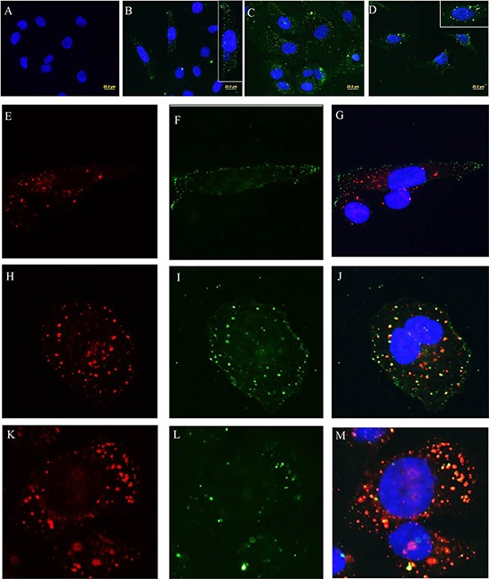
The metalloproteinase SAS1B [ovastacin, ASTL, astacin-like] was immunolocalized on the oolemma of ovulated human oocytes and in normal ovaries within the pool of growing oocytes where SAS1B protein was restricted to follicular stages spanning the primary-secondary follicle transition through ovulation. Gene-specific PCR and immunohistochemical studies revealed ASTL messages and SAS1B protein in both endometrioid [74%] and malignant mixed Mullerian tumors (MMMT) [87%] of the uterus. A MMMT-derived cell line, SNU539, expressed cell surface SAS1B that, after binding polyclonal antibodies, internalized into EEA1/LAMP1-positive early and late endosomes. Treatment of SNU539 cells with anti-SAS1B polyclonal antibodies caused growth arrest in the presence of active complement. A saporin-immunotoxin directed to SAS1B induced growth arrest and cell death. The oocyte restricted expression pattern of SAS1B among adult organs, cell-surface accessibility, internalization into the endocytic pathway, and tumor cell growth arrest induced by antibody-toxin conjugates suggest therapeutic approaches that would selectively target tumors while limiting adverse drug effects in healthy cells. The SAS1B metalloproteinase is proposed as a prototype cancer-oocyte tumor surface neoantigen for development of targeted immunotherapeutics with limited on-target/off tumor effects predicted to be restricted to the population of growing oocytes.

Keywords: ASTL/SAS1B/ovastacin; cancer surface neoantigen; immunotherapy target; oocyte-specific protein; uterine tumor biomarkers.


Conflict of interest statement

CONFLICTS OF INTEREST

The University of Virginia has filed patent applications on the use of the SAS1B as a cancer drug and diagnostic target. Neoantigenics has an exclusive license. E.P, J.H., A.M., K.A., and B.P. hold equity in Neoantigenics.

Figures

Figure 1
Figure 1. ASTL/SAS1B has ovary and oocyte restricted expression among normal human tissues, and a population of SAS1B is found on the oolemma of ovulated human oocytes
Panel A: RT-PCR analyses of 6 normal human ovarian cDNAs (H1-H6) using a c-terminus ASTL specific primer set show 309 base pair ASTL amplicons in each specimen. Panel B: ASTL expression was restricted to ovarian specimens (lane OVA) while other tissues (uterus, testes, placenta, adrenal, pancreas, spleen, kidney, liver and leukocytes) were ASTL silent. GAPDH was used as a loading control for all RT-PCR assays. Panels C and D: Immunohistochemical localization of SAS1B in human ovarian tissue sections with anti-SAS1B antibodies (C- IM antibodies and D1- PPpAb, D2- PIM antibodies/ normal rabbit IgG). SAS1B protein was localized to oocytes in primary-secondary transition follicles (C3), early antral follicles (C4) and beyond (D1). Oocytes within primordial follicles (C1) and small primary follicles i.e. resting follicles (C2) did not express SAS1B (green arrows). An early developing primary-secondary transition follicle (black arrow, C3, with development of a bilaminar layer noted at white arrow) shows initiation of SAS1B expression in the perinuclear region of the oocyte (inset) presumably in the Golgi apparatus. Panel E: Live stained fertilized human oocytes show membrane staining of SAS1B by immunofluorescence. Top inset shows phase image and bottom inset shows PIM control. Panel F: Western blots reveal a 46 kDa SAS1B protein (red asterisk) in human ovarian protein extracts (HO) with IM antibodies (F1, HO) and PPpAb (F2, HO). In protein extracts from normal human uterus (F1, HU and F2, HU) SAS1B was not detected.
Figure 2
Figure 2. SAS1B expression occurs at high incidence in uterine tumors
Panel A: Normal human uterine cDNA samples U1–U7 show no ASTL expression (lack of 309 base pair amplimer). Representative images indicating SAS1B expression in tumors; E1–7 (grade 1 endometrioid tumors) are ASTL positive, while in E8–11 (grade 3 endometrioid tumors) only E10 is positive. Panel B: In this group of 15 uterine tumors (E12–E26) 11 were positive for ASTL including 10 endometrioid specimens and one MMMT (E13). Panel C: cDNAs from myometrial tissue adjacent to the tumor (M12–26) show 5/15 positivity, likely due to tumor infiltration. Panel D: E27, 28, 30 and 32 are endometrioid tumors (4/4 positive) and E29 and 31 are MMMT (2/2 positive). Panel E: IHC using the IM or PIM (inset) antibodies on normal human proliferative (1) or secretory (2) endometrium showing absence of SAS1B protein. Immunohistochemical staining of SAS1B (brown DAB reaction product) in endometrioid cancers grade 1 (Panels 3–5), endometrioid cancers grade 3 (Panels 6–9), and MMMT's (Panels 10–13).
Figure 3
Figure 3. ASTL/SAS1B expression in MMMT derived cell lines
cDNAs from MMMT derived cell lines S08-38710 and SNU539 along with control MAD10 cells were probed (in duplicate) for ASTL using domain specific primers (Panels A, B, C represent ASTL amplicons of 237 base pair N-terminus, 309 base pair C-terminus and 579 base pair catalytic region, respectively). Panel D: ASTL transcription was noted in both S08-38710 and SNU539 while MAD10 was ASTL negative. GAPDH was used as a loading control. Panel F-H: On Western blots, major immunoreactive SAS1B protein isoforms (red asterisks, F) of ∼46 kDa [predicted from primary sequence] and ∼65 kDa, and a less abundant ∼36-37 kDa band, were identified in Celis extracts of SNU539 cells probed with IM antibodies, while blots exposed to PIM antibodies (E) were negative. MAD10 protein extracts showed no specific immunoreactivity with IM antibodies (H) or control PIM antibodies (G). In fixed S08-38710 (Panel I) and SNU539 (Panel J) cells IM antibodies localized SAS1B in the cytoplasm with high concentrations noted in the perinuclear region (Green is SAS1B, blue is DAPI nuclear counterstain, red is phalloidin counterstain for actin). No SAS1B immunoreactivity was noted in MAD10 cells (Panel K). Insets (I-K) show lack of immunoreactivity with PIM antibody on the respective cell types.
Figure 4
Figure 4. Membrane associated SAS1B in primary endometrioid cells and MMMT cell lines
Panel A: Live cell indirect immunofluorescence: Using IM antibodies, surface SAS1B green signals are seen in S08-38710 (A1), SNU539 (A2), luciferase expressing SNU539 (A3); red SAS1B signal is present in primary endometrioid tumor cells recovered from a patient (A4). Respective insets show no staining using PIM antibody. Panel B: Results of phase partitioning of SAS1B isoforms extracted with TX-114 from SNU539 cells where D: detergent phase; A: aqueous phase; P: pellet. Partitioning of various control proteins such as albumin in A phase (B1), GAPDH in D and A phases (B2), and CD44 in D phase (B3) was observed in their expected fractions. The PIM antibody stained no proteins (B4). The IM antibody showed the presence of the ∼36–37 kDa form predominantly in the D phase, the ∼65 kDa form was localized in the A phase and the 46 kDa form was predominantly seen in the A phase with a relatively small portion in the D phase. Un-partitioned 46 kDa form also appeared in the P phase (B5). The PPpAb detected the 46 kDa form in the A phase along with several other higher molecular weight proteins in the A and P phases (B6). Panel C: Recovery of endogenous SAS1B microsequences by immunoprecipitation from SNU539 cells. Immunoprecipitated proteins were obtained using IM antibody, run on 2-D gels, and Western blotted with IM antibodies (C1), ASTL PP antibody (C2), or PIM sera (C3). The immunoreactive spots (red circles) were detected with both anti-SAS1B antibody reagents (C1 & c2) ranging between pI 5-6 while the immunoprecipitate stained with the PIM antibody did not contain detectable SAS1B proteins (C3). The trail in the right side of the blots is the heavy chain of IgG detected by the secondary antibody. A duplicate immunoprecipitate was sent for mass spectrometry and the ASTL/SAS1B peptides shown in various colors were recovered (C4), confirming reactivity of the IM antibodies with endogenous SAS1B.
Figure 5
Figure 5. Complement mediated cytotoxicity with anti-SAS1B antibodies
Cells grown on impedance electrodes were used to study the effects of antibodies and complement on cell growth. Panels A–C show SNU539 cells, while D–F depict MAD10 cells. Panels A and D: These panels show effects of media alone, media with a control rabbit IgG, and Triton X-100 detergent on impedance. Cells incubated with detergent lost contact with electrodes resulting in decreased impedance thus indicating growth arrest. Panels B and E: These panels show treatment with IM and PIM antibodies in the presence of active rabbit complement proteins. SNU539 cells (B) lost contact with electrodes and showed a drop in impedance upon treatment with IM but not with PIM antibodies, while no such effects were seen with MAD10 cells (E). When the complement proteins were deactivated by heat in-activation at 56°C, no decreases in impedance were observed with the IM antibodies on SNU539 cells (Panel C) and MAD10 cells (Panel F). PIM antibodies in all conditions showed no decreases in impedance.
Figure 6
Figure 6. SAS1B-antibody complexes undergo endocytosis and co-localize with classical endocytic markers
At various time points after SAS1B antibody treatment cells were studied to evaluate intracellular entry and co-localization with markers of classical endocytic vesicles and lysosomes. Panel A: This panel shows no internalization with control PIM antibodies after 60 min, while IM antibodies at 15 min (Panel B), 30 min (Panel C) and 60 min (Panel D) show green immunofluorescent vesicles representing internalization of SAS1B-antibody complexes. Insets in B and D show higher magnifications. Punctuate green SAS1B+ endosomes were evident just beneath the cell membrane after 15 min of incubation at 37°C (Panel F) and did not co-localize with red EEA1+ vesicles (Panel E and merged in Panel G). As incubation periods increased SAS1B+ vesicles coalesced into larger vesicles deeper within the cytoplasm (Panels I and L). These larger SAS1B+ vesicles co-localized with red EEA1+ vesicles (Panel H and merged in Panel J) at 30 min and with red LAMP1+ vesicles (Panel K and merged in Panel M) at 60 min.
Figure 7
Figure 7. Arrest of SNU539 cell growth with a saporin-immunotoxin targeting SAS1B
Cells in culture were exposed to varying concentrations of IM and PIM antibodies complexed with a fixed concentration of 5.42 nM secondary saporin immunoconjugate and monitored over 72 hours. Cells were observed by light microscopy and assayed for total biomass; culture supernatants were assayed for LDH levels. Panel A: This shows mean percent survival of SNU539 cells in the presence of IM antibody saporin conjugates (N = 4 experiments). IM antibody at concentrations from 1 μM to 1 nM was used and concentrations of 1–10 nM showed significant inhibitory effects (7A and 7B) on growth while PIM antibodies at identical concentrations did not (blue bars 7A). Triton X-100 detergent was used as positive control to arrest growth at the outset of the treatment period (purple bar 7A). Normal rabbit IgG saporin, saporin conjugate alone (SCS), or media alone did not demonstrate growth arrest (7A). Panel B: Deleterious effects on cells noted by light microscopy include cell vacuolation, cell rounding, pyknosis, and death (7B9, magnified in 7B10). Panel C: Under identical conditions SAS1Bneg MAD10 cells did not exhibit growth arrest in culture (7C) and MAD10 cells did not demonstrate deleterious microscopic effects after similar treatments (Panel 7C1–7C3).

References

    1. Sachdev M, Mandal A, Mulders S, Digilio LC, Panneerdoss S, Suryavathi V, Pires E, Klotz KL, Hermens L, Herrero MB, Flickinger CJ, van Duin M, Herr JC. Oocyte specific oolemmal SAS1B involved in sperm binding through intra-acrosomal SLLP1 during fertilization. Developmental Biology. 2012;363:40–51. - PMC - PubMed
    1. Pires ES, Hlavin C, Macnamara E, Ishola-Gbenla K, Doerwaldt C, Chamberlain C, Klotz K, Herr AK, Khole A, Chertihin O, Curnow E, Feldman SH, Mandal A, et al. SAS1B protein [Ovastacin] shows temporal and spatial restriction to oocytes in several eutherian orders and initiates translation at the primary to secondary follicle transition. Developmental Dynamics. 2013;242:1405–26. - PMC - PubMed
    1. Burkart AD, Xiong B, Baibakov B, Jiménez-Movilla M, Dean J. Ovastacin, a cortical granule protease, cleaves ZP2 in the zona pellucida to prevent polyspermy. The Journal of Cell Biology. 2012;197:37–44. - PMC - PubMed
    1. Mouse EST dataset from GENBANK for Astl: http://www.ncbi.nlm.nih.gov/UniGene/ESTProfileViewer.cgi?uglist=Mm.31313.
    1. Website: human ASTL EST database, http://www.ncbi.nlm.nih.gov/nucest/14468989 and http://www.ncbi.nlm.nih.gov/UniGene/ESTProfileViewer.cgi?uglist=Hs.447993.

Publication types

MeSH terms