. 2015 Sep 1;10(9):e0135292.
doi: 10.1371/journal.pone.0135292. eCollection 2015.

Drug Development in Conformational Diseases: A Novel Family of Chemical Chaperones that Bind and Stabilise Several Polymorphic Amyloid Structures

Affiliations

Drug Development in Conformational Diseases: A Novel Family of Chemical Chaperones that Bind and Stabilise Several Polymorphic Amyloid Structures

Marquiza Sablón-Carrazana et al. PLoS One. .

Abstract

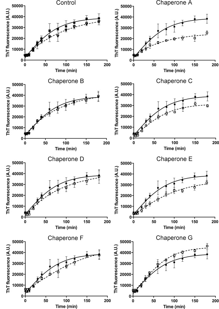
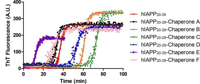
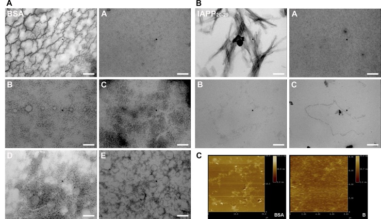
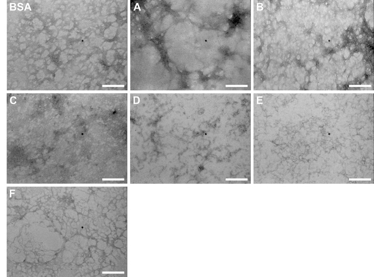
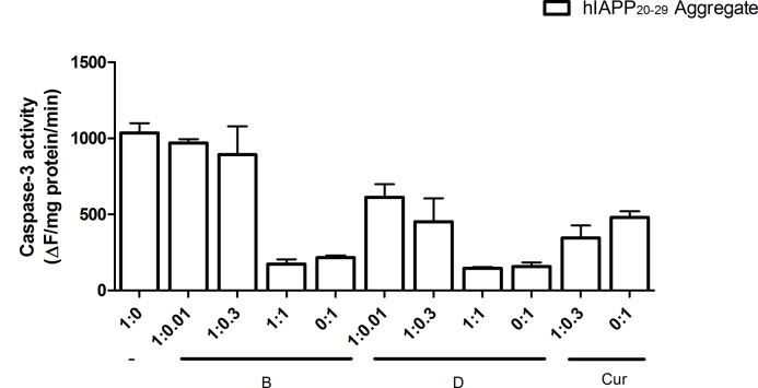
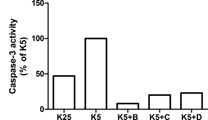
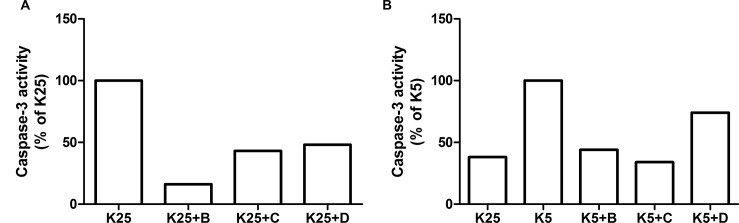
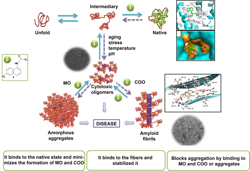
The increasing prevalence of conformational diseases, including Alzheimer's disease, type 2 Diabetes Mellitus and Cancer, poses a global challenge at many different levels. It has devastating effects on the sufferers as well as a tremendous economic impact on families and the health system. In this work, we apply a cross-functional approach that combines ideas, concepts and technologies from several disciplines in order to study, in silico and in vitro, the role of a novel chemical chaperones family (NCHCHF) in processes of protein aggregation in conformational diseases. Given that Serum Albumin (SA) is the most abundant protein in the blood of mammals, and Bovine Serum Albumin (BSA) is an off-the-shelf protein available in most labs around the world, we compared the ligandability of BSA:NCHCHF with the interaction sites in the Human Islet Amyloid Polypeptide (hIAPP):NCHCHF, and in the amyloid pharmacophore fragments (Aβ17-42 and Aβ16-21):NCHCHF. We posit that the merging of this interaction sites is a meta-structure of pharmacophore which allows the development of chaperones that can prevent protein aggregation at various states from: stabilizing the native state to destabilizing oligomeric state and protofilament. Furthermore to stabilize fibrillar structures, thus decreasing the amount of toxic oligomers in solution, as is the case with the NCHCHF. The paper demonstrates how a set of NCHCHF can be used for studying and potentially treating the various physiopathological stages of a conformational disease. For instance, when dealing with an acute phase of cytotoxicity, what is needed is the recruitment of cytotoxic oligomers, thus chaperone F, which accelerates fiber formation, would be very useful; whereas in a chronic stage it is better to have chaperones A, B, C, and D, which stabilize the native and fibril structures halting self-catalysis and the creation of cytotoxic oligomers as a consequence of fiber formation. Furthermore, all the chaperones are able to protect and recondition the cerebellar granule cells (CGC) from the cytotoxicity produced by the hIAPP20-29 fragment or by a low potassium medium, regardless of their capacity for accelerating or inhibiting in vitro formation of fibers. In vivo animal experiments are required to study the impact of chemical chaperones in cognitive and metabolic syndromes.


Conflict of interest statement

Competing Interests: The authors of this manuscript have the following competing interests: The author declares having filed two patent applications, WO2010118706 A2 and WO2014131374 A1. This does not alter the authors' adherence to PLOS ONE policies on sharing data and materials.

Figures

Fig 1
Fig 1. Structures and LogP of the chemical chaperones.
N-(2-aminoethyl)-N'-1-naphthylsuccinamide A; methyl (2-{[4-(1-naphthylamino)-4-oxobutanoyl]amino}ethyl) dithiocarbamate B; (2R)-2-(6-methoxy-2-naphthyl)propanoic acid (Naproxen) C; N-[4-(1-naphthylamino)-4-oxobutanoyl]-β-alanine D; 6-{[4-(1-naphthylamino)-4-oxobutanoyl]amino} hexanoic acid E; N 3,N 3'-ethane-1,2-dyilbis(N 1-1-naphthylsuccinamide) F; N-(4-aminobutyl)-N'-1-naphthylsuccinamide G; (1E,6E)-1,8-bis(4-hydroxy-3-methoxyphenyl)octa-1,6-diene-3,5-dione (Curcumine) H; 7-hydroxy-8-[(Z)-phenyldiazenyl]naphthalene-1,3-disulfonic acid (Orange G) I; (2Z)-3-[6-(dimethylamino)-2-naphthyl]-2-isocyanobut-2-enenitrile (DDNP) J. *The logP was calculated using ACD/Log P software (ACD/1-Lab Service, Toronto, Ontario, Canada). ** Value estimated for uncharged compound.
Fig 2
Fig 2. Molecular docked model of chemical chaperones (stick representation), located within the hydrophobic pocket of BSA (cartoon and sphere views).
At 5Å distance, the amino acid residues surrounding chaperones (A, B, C, D, E, F, I and H) are represented in orange. The H-bonding interaction between chaperones and amino acid residues of BSA, are shown as a dotted line.
Fig 3
Fig 3. Visualization of binding site of chaperones (A, B, C, D, E, F, G and H) into IIA and IIIA regions of BSA, within 5Å distance.
Fig 4
Fig 4. Molecular docked model of chemical chaperones (stick representation), located in the amino acids of Aβ17–42 segment (cartoon and sphere views).
At 5Å distance, the amino acid residues surrounding chaperonins (A, B, C, D, E, F, H, I and J) are represented in orange. The hydrogen bonding interaction between chaperones and amino acid residues of Aβ17–42 segment are shown as a dotted line.
Fig 5
Fig 5. Meta-structure of pharmacophore.
A structural alignment using Pymol, whereby the interaction zones between β-amiloyd17–42 (Aβ17–42), IAPP and Eisenberg’s pharmacophore molecules and the chaperons were set in position with the interaction zone between BSA and the chaperons. BSA is shown in white at 70% transparency, the interaction zone between the Aβ17–42 and the chaperons is shown in red, the one corresponding to the Eisenberg pharmacophore in blue, with IAPP in yellow and with BSA in green.
Fig 6
Fig 6. A. ThT fluorescence kinetics during amyloid fibrillation of BSA.
Experiments were carried out at 65°C in glycine buffer (pH = 3; 0.05M, NaCl 100 mM), in presence or absence of the selected chaperones at molar relation 1:1 of BSA: Chap. (ThT: 24 μM). Time dependent changes in ThT intensity was fitted by exponential function (solid line). In all cases, the adjusted R-square values were greater than 0.9. The experiments were carried out in triplicate. The control BSA alone (black square) and BSA plus DMSO (white triangles). All the chaperones (empty squares) were compared with BSA plus DMSO (black squares). B: Normalized maximum values of the ThT fluorescence intensity of BSA fibrilations in presence or absence of chaperones (A, B, C, D, E, F and G).
Fig 7
Fig 7. ThT fluorescence kinetics during amyloid fibrillation of IAPP20–29 (100 μmol/L).
Experiments were carried out at 25°C in PBS buffer (pH 7.4; 10 mmol/L; NaCl 100 mmol/L), in presence or absence of the selected chaperones at molar relation 1:1 of IAPP20–29: Chap. (ThT: 24 μM). Time dependent changes in ThT intensity was fitted by sigmoidal function (solid line). The experiments were carried out in triplicate. tlag of fibril formation of the tested chaperones * p < 0,05 show significant differences with regard to control.
Fig 8
Fig 8. A. Transmission Electron Microscopy (TEM) micrographs of BSA fibrilogenesis process (75 μmol/L), with and without N-(2-aminoethyl)-N'-1-naphthylsuccinamide
A; methyl (2-{[4-(1-naphthylamino)-4-oxobutanoyl]amino}ethyl) dithiocarbamate B; (2R)-2-(6-methoxy-2-naphthyl)propanoic acid (Naproxen) C; N-[4-(1-naphthylamino)-4-oxobutanoyl]-β-alanine D and 6-{[4-(1-naphthylamino)-4-oxobutanoyl]amino} hexanoic acid E, at 70°C. B: Transmission Electron Microscopy (TEM) micrographs of hIAPP20–29 fibrilogenesis process with and without N-(2-aminoethyl)-N'-1-naphthylsuccinamide A; methyl (2-{[4-(1-naphthylamino)-4-oxobutanoyl] amino} ethyl) dithiocarbamate B and (2R)-2-(6-methoxy-2-naphthyl)propanoic acid (Naproxen) C; at 25°C. Molar ratio hIAPP20–29: chaperones was 1:1. C: Atomic Force Microscopy (AFM) micrographs of BSA fibrilogenesis process (75 μmol/L), with and without B, at 70°C. Molar ratio BSA:chaperones was 1:1.
Fig 9
Fig 9. Transmission Electron Microscope (TEM) images of the BSA fibril formed with or without the chaperones (A, B, C, D, E, F and G) at a molar relation of BSA: chaperones 1:1, in buffer Tris (pH = 7.4, 20 mM) and 75°C.
Scale bar 200 nm.
Fig 10
Fig 10. Cell Viability of CGC exposed to hIAPP20–29, in monomeric and aggregated form, through MTT assay, with and without N-[4-(1-naphthylamino)-4-oxobutanoyl]-β-alanine D; methyl (2-{[4-(1-naphthylamino)-4-oxobutanoyl]amino}ethyl) dithiocarbamate B and H used as reference; at different molar ratio hIAPP20–29:chaperones.
All the experiments have the same concentration of hIAPP20–29
Fig 11
Fig 11. Cell Apoptosis of CGC exposed to hIAPP20–29, in monomeric and aggregated form, with and without N-[4-(1-naphthylamino)-4-oxobutanoyl]-β-alanine D; methyl (2-{[4-(1-naphthylamino)-4-oxobutanoyl]amino}ethyl) dithiocarbamate B and H used as reference; at different molar ratio hIAPP20–29:chaperones.
In all assays caspase-3 levels were measured by immunofluorescence.
Fig 12
Fig 12. Cell Apoptosis of CGC exposed to pro-apoptotic stimulus as low potassium concentration (K5) in the presence of N-[4-(1-naphthylamino)-4-oxobutanoyl]-β-alanine D; methyl (2-{[4-(1-naphthylamino)-4-oxobutanoyl] amino}ethyl) dithiocarbamate B and C used as reference.
In all assays caspase-3 levels were measured by inmunofluorescence.
Fig 13
Fig 13. Cell Apoptosis of CGC exposed to pro-apoptotic stimulus as low potassium concentration (K5) during 4 h and later, the chaperones N-[4-(1-naphthylamino)-4-oxobutanoyl]-β-alanine D; methyl (2-{[4-(1-naphthylamino)-4-oxobutanoyl]amino}ethyl) dithiocarbamate B and C (reference), were inoculated.
In all assays caspase-3 levels were measured by inmunofluorescence.
Fig 14
Fig 14. Proposal mechanism of chaperone on protein aggregation/disaggregation.
Novel chemical chaperones family can regulate fiber formation processes by binding to the native state, minimizing the formation of amorphous aggregates, as well as cytotoxic oligomers.

Similar articles

Cited by

References

    1. Gidalevitz T, Ben-Zvi A, Ho KH, Brignull HR, Morimoto RI (2006) Progressive disruption of cellular protein folding in models of polyglutamine diseases. Science 311: 1471–1474. 1124514 [pii];10.1126/science.1124514 - DOI - PubMed
    1. Landau M, Sawaya MR, Faull KF, Laganowsky A, Jiang L, Sievers SA, et al. (2011) Towards a pharmacophore for amyloid. PLOS Biol 9: e1001080 10.1371/journal.pbio.1001080;PBIOLOGY-D-11-00136 [pii]. - DOI - PMC - PubMed
    1. Welch WJ (2004) Role of quality control pathways in human diseases involving protein misfolding. Semin Cell Dev Biol 15: 31–38. 10.1016/j.semcdb.2003.12.011;S1084952103001162 [pii]. - DOI - PubMed
    1. Westermark P, Benson MD, Buxbaum JN, Cohen AS, Frangione B, Ikeda S, et al. (2007) A primer of amyloid nomenclature. Amyloid 14: 179–183. 781269331 [pii];10.1080/13506120701460923 - DOI - PubMed
    1. Herczenik E, Gebbink MF (2008) Molecular and cellular aspects of protein misfolding and disease. FASEB J 22: 2115–2133. fj.07-099671 [pii];10.1096/fj.07-099671 - DOI - PubMed

Publication types

MeSH terms

LinkOut - more resources