. 2015 Dec;27(12):2444-51.
doi: 10.1016/j.cellsig.2015.08.017. Epub 2015 Aug 29.

PAQR3 regulates Golgi vesicle fission and transport via the Gβγ-PKD signaling pathway

Affiliations

PAQR3 regulates Golgi vesicle fission and transport via the Gβγ-PKD signaling pathway

Thamara Hewavitharana et al. Cell Signal. 2015 Dec.

Abstract

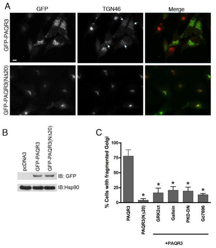
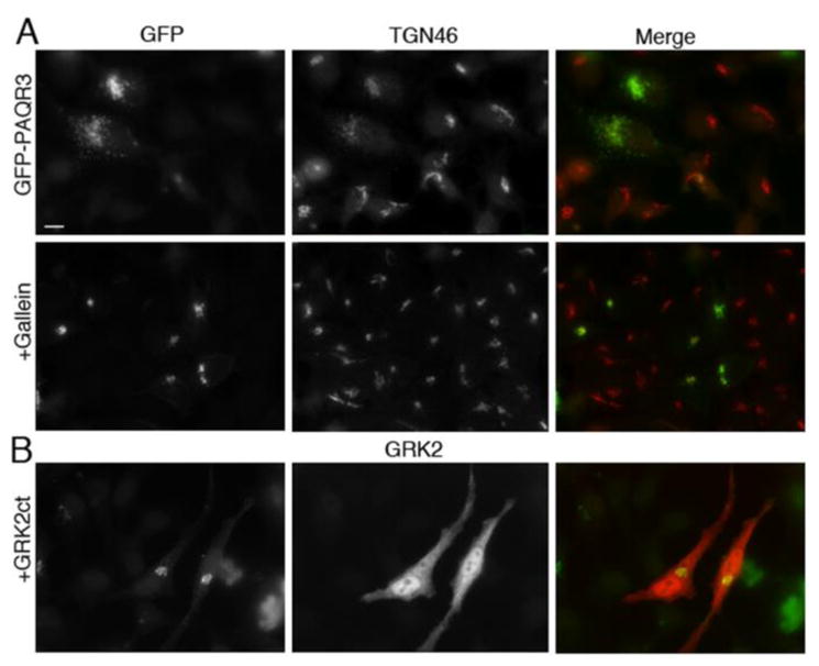
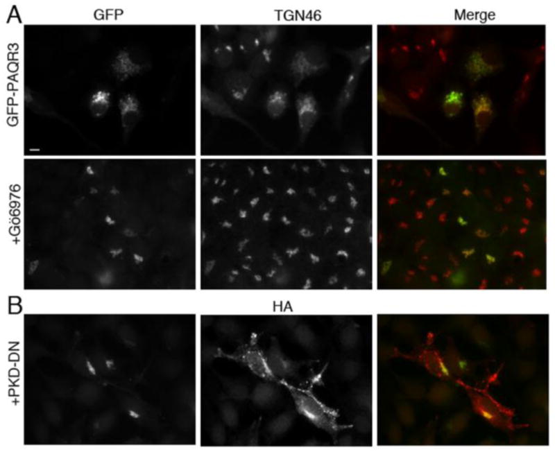
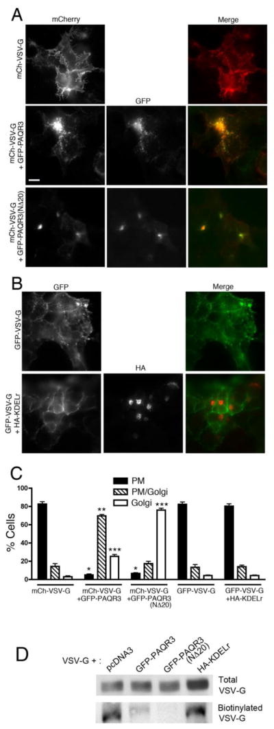
Heterotrimeric G proteins function at diverse subcellular locations, in addition to canonical signaling at the plasma membrane (PM). Gβγ signals at the Golgi, via protein kinase D (PKD), to regulate fission of PM-destined vesicles. However, the mechanism by which Gβγ is regulated at the Golgi in this process remains elusive. Recent studies have revealed that PAQR3 (Progestin and AdipoQ Receptor 3), also called RKTG (Raf Kinase Trapping to the Golgi), interacts with the Gβ subunit and localizes Gβ to the Golgi thereby inhibiting Gβγ signaling at the PM. Herein we show that, in contrast to this inhibition of canonical Gβγ signaling at the PM, PAQR3 promotes Gβγ signaling at the Golgi. Expression of PAQR3 causes fragmentation of the Golgi, while a Gβ binding-deficient mutant of PAQR3 does not cause Golgi fragmentation. Also, a C-terminal fragment of GRK2 (GRK2ct), which interacts with Gβγ and inhibits Gβγ signaling, and gallein, a small molecule inhibitor of Gβγ, are both able to inhibit PAQR3-mediated Golgi fragmentation. Furthermore, a dominant negative form of PKD (PKD-DN) and a pharmacological inhibitor of PKD, Gö6976, also inhibit PAQR3-mediated fragmentation of the Golgi. Importantly, expression of the Gβ binding-deficient mutant of PAQR3 inhibits the constitutive transport of the model cargo protein VSV-G from the Golgi to the PM, indicating the involvement of PAQR3 in Golgi-to PM vesicle transport and a dominant negative role for this mutant. Collectively, these results reveal a novel role for the newly characterized, Golgi-localized PAQR3 in regulating Gβγ at the non-canonical subcellular location of the Golgi and thus for controlling Golgi-to-PM protein transport via the Gβγ-PKD signaling pathway.

Keywords: Golgi; Heterotrimeric G protein; Membrane transport; Non-canonical signaling; Subcellular localization; Vesicle trafficking.


Figures

Figure 1
Figure 1. Expression of PAQR3 induces Golgi fragmentation while the Gβ binding-deficient mutant PAQR3(NΔ20) does not fragment the Golgi
A, HeLa cells were transfected with GFP-PAQR3 (top row) or GFP-PAQR3(NΔ20) (bottom row) for 48 h. Cells were fixed and processed for immunofluorescence microscopy using anti-TGN46 to detect Golgi morphology, and expressed PAQR3 and PAQR3(NΔ20) were detected by intrinsic GFP fluorescence. Arrows indicate Golgi fragmentation. Bar, 10 μm. B, To confirm equivalent expression of GFP-PAQR3 and GFP-PAQR3(NΔ20), immunoblotting was performed using HeLa cell lysates after transfection with pcDNA3, GFP-PAQR3 or GFP-PAQR3(NΔ20). An anti-GFP antibody was used to detect GFP-PAQR3 or GFP-PAQR3(NΔ20) (upper panel), and an anti-Hsp90 antibody was used as a gel loading control (lower panel). Consistent with the immunoblot shown, previous results have demonstrated that PAQR3 and PAQR3(NΔ20) display no detectable difference in mobility [24, 62]. C, Shown is a bar graph quantitating the effect of PAQR3, PAQR3(NΔ20), and the effect of Gβγ and PKD inhibitors in the presence of PAQR3 on Golgi fragmentation. Cells were transfected/treated as described in Figure Legends 1A, 2 and 3. Values are the means +/− S.D. for 3 separate experiments. 100 cells were counted in each experiment. Asterisks indicate statistical significance (p<0.001, t-test) compared to PAQR3 (first bar).
Figure 2
Figure 2. Gβγ inhibitors GRK2ct and gallein inhibit PAQR3-mediated Golgi fragmentation
A, HeLa cells were transfected with GFP-PAQR3 for 36–48 h and then treated with 10 μM gallein (bottom row) or vehicle (top row) for 8 h prior to being fixed and processed for immunofluorescence microscopy using anti-TGN46 to detect Golgi morphology. Expressed GFP-PAQR3 was detected by intrinsic GFP fluorescence. B, HeLa cells were transfected with GFP-PAQR3 together with GRK2ct. Cells were fixed and stained with anti-GRK2, and the intrinsic GFP fluorescence allowed visualization of GFP-PAQR3, and, by extension, Golgi morphology. Bar, 10 μm.
Figure 3
Figure 3. PKD inhibitors PKD-DN and Gö6976 inhibit PAQR3-mediated Golgi fragmentation
A, HeLa cells were transfected with GFP-PAQR3 for 36–48 h and then treated with 5 μM Gö6976 (bottom row) or vehicle (top row) for 8 h prior to being fixed and processed for immunofluorescence microscopy using anti-TGN46 to detect Golgi morphology. Expressed GFP-PAQR3 was detected by intrinsic GFP fluorescence. B, HeLa cells were transfected with GFP-PAQR3 together with HA-PKD-DN (PKD-K618N). Cells were fixed and stained with anti-HA, and the intrinsic GFP fluorescence allowed visualization of GFP-PAQR3, and, by extension, Golgi morphology. Bar, 10 μm.
Figure 4
Figure 4. Gβ binding-deficient mutant PAQR3(NΔ20) inhibits VSV-G transport from the Golgi to the PM
A, HEK293 cells were transfected with mCherry-VSV-G alone (top row) or together with GFP-PAQR3 (middle row) or GFP-PAQR3(NΔ20) (bottom row), incubated for 24 h and then transferred to the non-permissive temperature of 39 °C for 16 h. Cells were then shifted to 20 °C for 2 h (after 1 h of which cyclohexamide was added) at which temperature VSV-G localizes to and is retained at the Golgi. Cells were then transferred to the permissive temperature of 32 °C for 2 h for VSV-G transport to the PM. Cells were then fixed and stained with anti-VSV-G followed by Alexa 594-conjugated secondary antibody to better visualize mCherry-VSV-G. PAQR3 and PAQR3-(NΔ20) were detected by intrinsic GFP fluorescence. Bar, 10 μm. B, HEK293 cells were transfected with GFP-VSV-G alone (top row) or together with HA-KDELr (bottom row) and the VSV-G transport assay was performed as described in A. After 2 h incubation at the permissive temperature of 32 °C, cells were fixed and stained with anti-HA antibody to visualize KDELr. GFP-VSV-G was visualized by intrinsic GFP fluorescence. C, Shown is a bar graph quantitating the localization of VSV-G in the absence or presence of co-expressed PAQR3, PAQR3(NΔ20) or KDELr. VSV-G localization in individual cells was scored as predominantly PM (PM), both PM and Golgi (PM/Golgi), or predominantly Golgi (Golgi). Values depicted are the means (+/− S.D., vertical bars) for 3 separate experiments. 100 cells were counted in each experiment. Asterisks indicate statistical significance (p<0.001, t-test) of the indicated bar (*, PM; **, PM/Golgi; ***, Golgi) compared to the corresponding bar of the mCh-VSV-G only sample (first set of bars). D, HEK293 cells were transfected with mCherry-VSV-G together with pcDNA3, GFP-PAQR3, GFP-PAQR3(NΔ20) or HA-KDELr, as indicated, and cells were cultured as described above for A and B. After 2 h at 32 °C to allow transport of VSV-G to the PM, cell surface proteins were biotinylated and isolated as described under Materials and Methods. VSV-G in total cell lysates (upper panel) and cell surface biotinylated VSV-G (lower panel) were detected by immunoblotting with an anti-VSV-G antibody

Similar articles

Cited by

References

    1. Oldham WM, Hamm HE. Nat Rev Mol Cell Biol. 2008;9(1):60–71. - PubMed
    1. Milligan G, Kostenis E. Br J Pharmacol. 2006;147(Suppl 1):S46–55. - PMC - PubMed
    1. Hewavitharana T, Wedegaertner PB. Cell Signal. 2012;24(1):25–34. - PMC - PubMed
    1. Boularan C, Kehrl JH. Cell Signal. 2014;26(6):1269–1282. - PMC - PubMed
    1. Ahmed SM, Angers S. J Recept Signal Transduct Res. 2013;33(3):177–183. - PubMed

Publication types

MeSH terms

Substances