Review
. 2015 Sep;32(3):278-88.
doi: 10.1055/s-0035-1556826.

Current Status of Percutaneous Endografting

Affiliations
Review

Current Status of Percutaneous Endografting

Parag J Patel et al. Semin Intervent Radiol. 2015 Sep.

Abstract

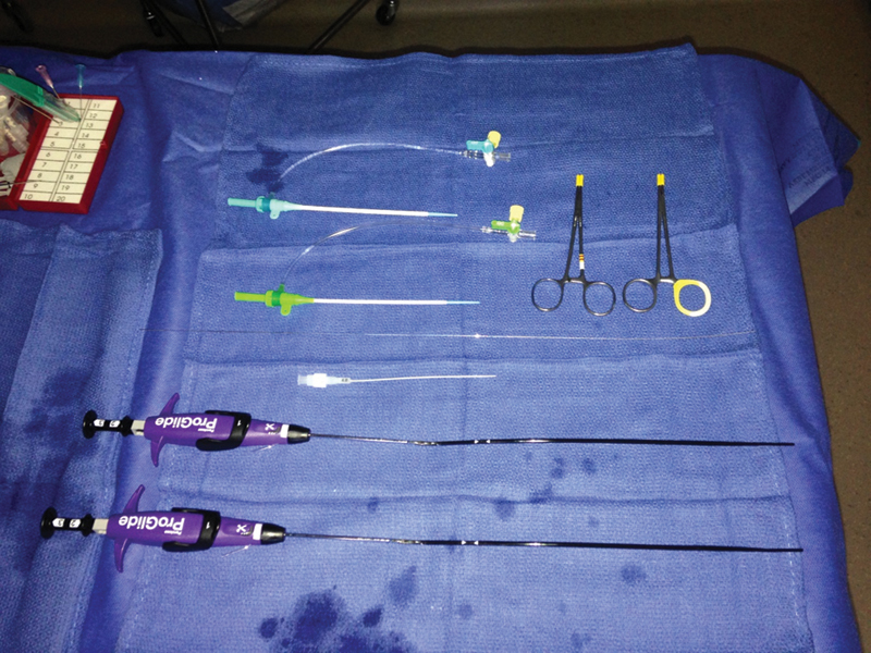
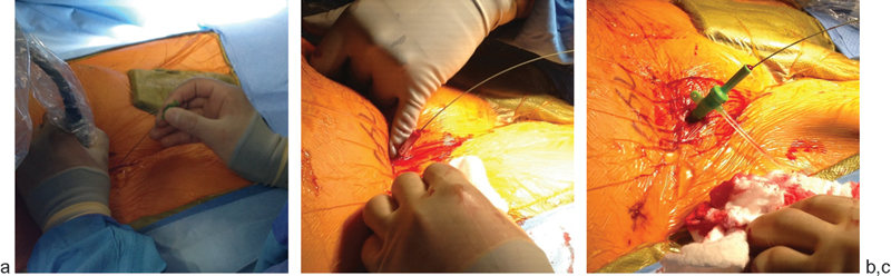
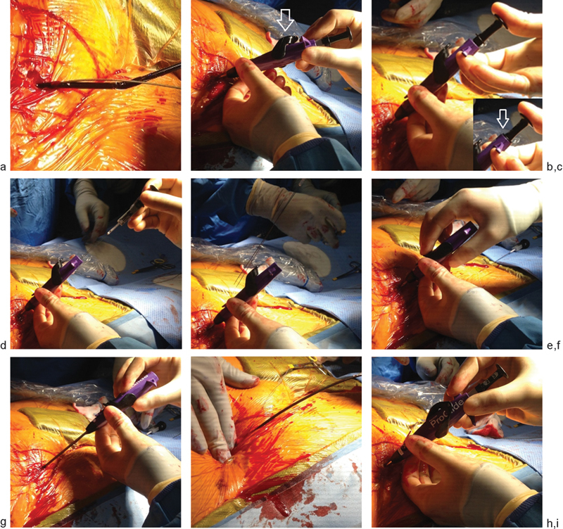
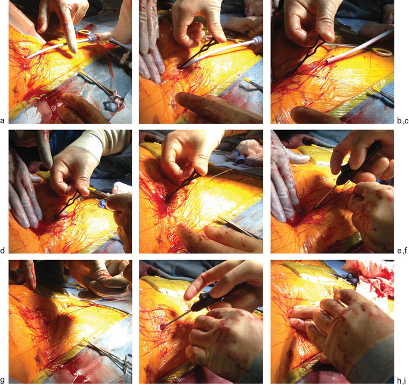
Totally percutaneous endovascular abdominal aortic aneurysm repair (PEVAR) using suture-mediated closure devices (SMCDs) has several well-established advantages over standard open femoral exposure as a direct consequence of being less invasive and having shorter times to hemostasis and procedure completion. The first multicenter randomized controlled trial designed to assess the safety and efficacy of PEVAR and to compare percutaneous access with standard open femoral exposure was recently published (the PEVAR trial). The PEVAR trial demonstrated that percutaneous endografting is safe, effective, and noninferior to standard open femoral exposure among trained operators. The study reaffirmed the results of several recent single center and nonrandomized studies, demonstrating that percutaneous access facilitated shorter procedures, shorter times to secure hemostasis, and improved quality of life for patients. As PEVAR has gained popularity among patients and physicians, refinements to the technique and patient selection process have been made. There has been growing interest in treating patients with anatomical characteristics previously thought to be unsuitable for PEVAR, such as common femoral artery (CFA) calcifications, scarred groins, small CFA diameter, and high patient body mass index (BMI). However, observance of strict procedural technique and consideration for patient selection criteria remain paramount in achieving acceptable technical success rates with PEVAR.

Keywords: PEVAR; ProGlide; interventional radiology; preclose.


Figures

Fig. 1
Fig. 1
Materials necessary for percutaneous suture mediated closure: 6F sheath; 8F sheath. Micropuncture wire and transitional dilator. Two ProGlide devices. Two suture tags.
Fig. 2
Fig. 2
(a) Access the common femoral artery with a micropuncture needle under ultrasound guidance at the 12-o'clock position of the vessel. (b) Dissect the subcutaneous tissues down to the anterior wall of the common femoral artery using blunt technique, shown here using a finger. (c) Predilate the tract and arteriotomy with a 6F sheath over a 0.035-in. guidewire.
Fig. 3
Fig. 3
(a) Advance the first Perclose ProGlide device over the guidewire until the guide wire exit port is just above the skin line. Remove the guidewire and then advance the device at a 45-degree angle until brisk, pulsatile blood flow is seen at the clear plastic marker lumen on the right side of the device. (b) Rotate the device to the medial position while maintaining a 45-degree angle and deploy the footplate using the black lever marked #1 on the device (shown here with the footplate deployed). (c) Apply gentle backward force to the device to bring the footplate into contact with the vessel wall. Deploy the needle by pushing on the plunger in the direction of the arrow marked #2 on the device until the black collar of the plunger makes contact with the purple body of the device (image inset). (d) Maintain gentle retraction on the device to stabilize it as the suture is retrieved by retracting the plunger marked #3 on the device to disengage the suture needles. (e) Continue pulling back on the plunger until the suture is taut, confirming that the entirety of suture has been retracted from the body of the device. Use the quick cut mechanism on the device to cut the suture from the needle. (f) Push the lever (marked on its undersurface with #4) on the device to return the footplate to the closed position after relaxing retraction on the device. (g) Gently withdraw the device until the guide wire exit port is visible at the skin line. Grasp the two suture limbs adjacent to the sheath and pull the suture limbs through the proximal guide. Re-advance the 0.035-in. guidewire through the guidewire exit port. (h) As the first Perclose ProGlide device is being removed, maintain manual pressure over the arteriotomy site to prevent formation of a hematoma. (i) Steps are repeated with the second Perclose ProGlide device delivered in the lateral position. Once completed, set aside both pairs of sutures under a damp towel during the EVAR procedure.
Fig. 4
Fig. 4
(a) Irrigate the sutures to remove debris or dried tissue and blood before advancing the knots. (b) The knot pusher is placed on the medial rail suture. The thumb is positioned at the top of the knot pusher. The suture should be aligned coaxial with the tissue tract. (c) Apply gentle manual pressure to the proximal CFA to maintain hemostasis as the sheath is removed over a guidewire. (d) Tighten the knot by pulling the shorter, white tipped suture limb, keeping it coaxial to the tissue tract. (e) While maintaining gentle pressure on the knot pusher and the suture limbs taut, manual pressure over the common femoral artery is removed to assess the degree of hemostasis. (f) Use the red lever on the suture trimmer to cut the suture limbs. Manual pressure is applied over the common femoral artery as the suture limbs are cut. Note that the 0.035-in. guidewire remains in position. (g) Repeat with the second set of suture limbs. The snared knot pusher is used to approximate the tissue edges. With the guidewire still in position, pull up on rail suture, advancing the knot down to the arteriotomy. With gentle traction held on the rail suture, manual compression is released from the proximal CFA to assess for hemostasis (shown). (h) Following guidewire removal, the knot is locked and then the second set of suture limbs are cut and removed. (i) Manual pressure should be held for approximately 10 minutes.

Similar articles

Cited by

References

    1. Nelson P R, Kracjer Z, Kansal N. et al.A multicenter, randomized, controlled trial of totally percutaneous access versus open femoral exposure for endovascular aortic aneurysm repair (the PEVAR trial) J Vasc Surg. 2014;59(5):1181–1193. - PubMed
    1. Haas P C, Krajcer Z, Diethrich E B. Closure of large percutaneous access sites using the Prostar XL Percutaneous Vascular Surgery device. J Endovasc Surg. 1999;6(2):168–170. - PubMed
    1. Dosluoglu H H, Cherr G S, Harris L M, Dryjski M L. Total percutaneous endovascular repair of abdominal aortic aneurysms using Perclose ProGlide closure devices. J Endovasc Ther. 2007;14(2):184–188. - PubMed
    1. Torsello G B, Kasprzak B, Klenk E, Tessarek J, Osada N, Torsello G F. Endovascular suture versus cutdown for endovascular aneurysm repair: a prospective randomized pilot study. J Vasc Surg. 2003;38(1):78–82. - PubMed
    1. Arslan B, Turba U C, Sabri S, Angle J F, Matsumoto A H. Current status of percutaneous endografting. Semin Intervent Radiol. 2009;26(1):67–73. - PMC - PubMed