

Massive peritoneal cavity calcification in the course of advanced ovarian cancer: a case report
- PMID: 26327904
- PMCID: PMC4498033
- DOI: 10.5114/pm.2015.52156
Massive peritoneal cavity calcification in the course of advanced ovarian cancer: a case report
Abstract
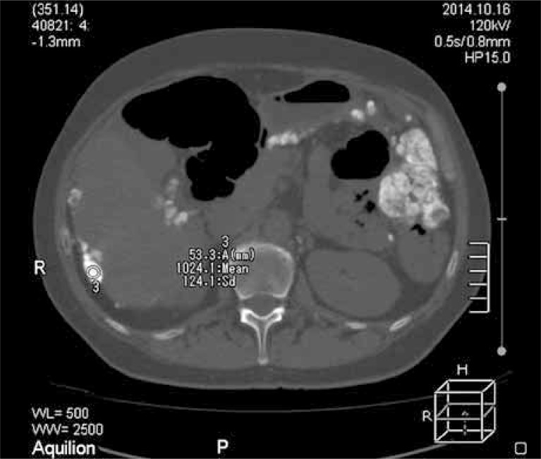
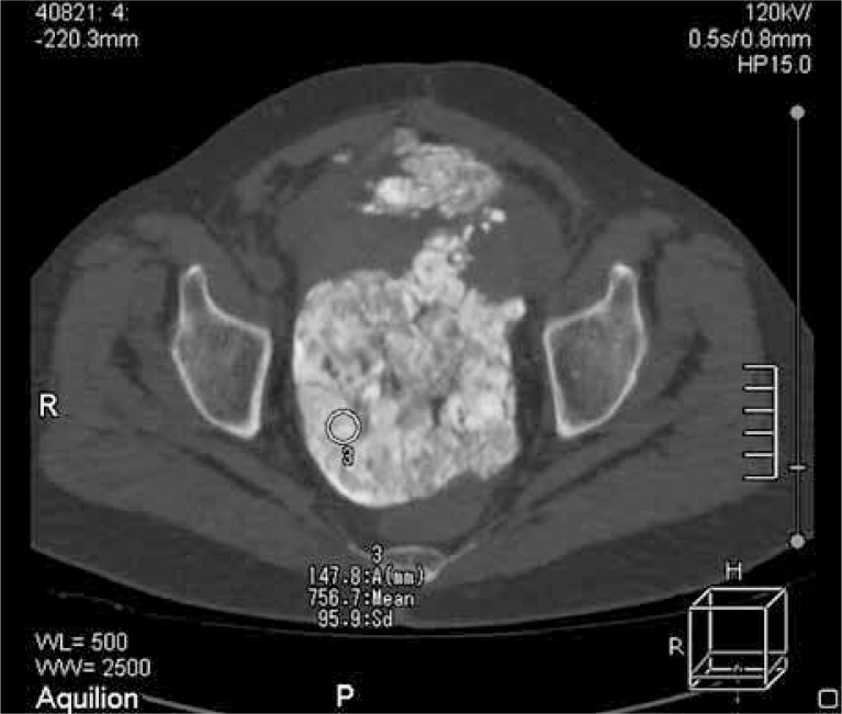
Ovarian cancer usually does not give any clinical signs until it reaches a large size. This condition is often associated with the occurrence of metastases within the peritoneal cavity, pelvic and abdominal cavities. Ovarian cancer can spread by intraperitoneal implantation, by way of the lymphatic system, and also through the systemic circulation. Even when the tumor reaches a large size, the symptoms are not specific and may resemble other ailments. Therefore, ovarian cancer is detected in most cases only in the third and fourth level of advancement. Peritoneal calcification occurs in many diseases. The degree of calcium deposits is usually small and does not give clinical symptoms. In the reported case, computed tomography of the abdomen showed numerous scattered peritoneal calcifications of irregular shape as well as massive calcification in the uterus and appendages. In the detection of changes associated with calcification, multidetectory computed tomography shows a very high sensitivity. It makes the precise location and assessment of the extent of changes possible.
Keywords: abdominal cavity; calcification peritoneum; ovarian cancer; peritoneal malignancy.
Figures
Similar articles
-
Localization and extent of peritoneal calcification in three uremic patients on continuous ambulatory peritoneal dialysis.Ther Apher Dial. 2008 Oct;12(5):413-6. doi: 10.1111/j.1744-9987.2008.00620.x. Ther Apher Dial. 2008. PMID: 18937728
-
Total parietal peritonectomy with en bloc pelvic resection for advanced ovarian cancer with peritoneal carcinomatosis.Gynecol Oncol. 2016 Dec;143(3):688-689. doi: 10.1016/j.ygyno.2016.10.014. Epub 2016 Oct 13. Gynecol Oncol. 2016. PMID: 27743737
-
A Way to Reduce the Occurrence of Intraoperative Capsule Rupture in Presumed Clinically Early-stage Ovarian Cancer with Adhesions to the Abdominal Wall.J Minim Invasive Gynecol. 2022 Jan;29(1):16. doi: 10.1016/j.jmig.2021.07.003. Epub 2021 Jul 12. J Minim Invasive Gynecol. 2022. PMID: 34265440
-
Imaging of peritoneal deposits in ovarian cancer: A pictorial review.World J Radiol. 2016 May 28;8(5):513-7. doi: 10.4329/wjr.v8.i5.513. World J Radiol. 2016. PMID: 27247717 Free PMC article. Review.
-
Role of ultrasound in advanced peritoneal malignancies.Minerva Med. 2019 Aug;110(4):292-300. doi: 10.23736/S0026-4806.19.06103-2. Epub 2019 May 6. Minerva Med. 2019. PMID: 31081311 Review.
Cited by
-
Well-Differentiated Papillary Mesothelioma with Omental Calcifications: A Case Report and Review of the Literature.Am J Case Rep. 2020 Jan 13;21:e920487. doi: 10.12659/AJCR.920487. Am J Case Rep. 2020. PMID: 31929500 Free PMC article.
References
-
- Urban A, Miszczyk L. Rak jajnika – diagnostyczny i terapeutyczny problem ginekologii onkologicznej. Contemp Oncol (Pozn) 2003;7:294–300.
-
- Whittemore AS, Harris R, Itnyre J. Characteristics relating to ovarian cancer risk: collaborative analysis of 12 US case-control studies, II. Invasive epithelial ovarian cancers in white women. Collaborative Ovarian Cancer Group. Am J Epidemiol. 1992;136:1184–1203. - PubMed
-
- Kim CK, Park BK, Choi JY, et al. Detection of recurrent ovarian cancer at MRI: comparison with integrated PET/CT. J Comput Assist Tomogr. 2007;31:868–875. - PubMed
-
- Kawamoto S, Urban BA, Fishman EK. CT of epithelial ovarian tumors. Radiographics. 1999;19:85–102. - PubMed
Publication types
LinkOut - more resources
Full Text Sources
Other Literature Sources