. 2015 Jul 15;7(7):1214-26.
eCollection 2015.

Transplantation of bradykinin-preconditioned human endothelial progenitor cells improves cardiac function via enhanced Akt/eNOS phosphorylation and angiogenesis

Affiliations

Transplantation of bradykinin-preconditioned human endothelial progenitor cells improves cardiac function via enhanced Akt/eNOS phosphorylation and angiogenesis

Zu-Long Sheng et al. Am J Transl Res. .

Abstract

This study determines whether preconditioning (PC) of human endothelial progenitor cells (hEPCs) with bradykinin promotes infarcted myocardium repair via enhanced activation of B2 receptor (B2R)-dependent Akt/eNOS and increased angiogenesis. hEPCs with or without bradykinin preconditioning (BK-PC) were transplanted into a nude mouse model of acute myocardial infarction. Survival of transplanted cells was assessed using DiD-labeled hEPCs. Infarct size, cardiac function, and angiogenesis were measured 10 d after transplantation. Akt, eNOS, and vascular endothelial growth factor (VEGF) expressions in cardiac tissues were detected by western blotting, and NO production was determined using an NO assay kit. The cell migration and tube formation in cultured hEPCs were determined using transwell chamber and matrigel tube formation assays, respectively. The VEGF levels in the cell supernatant were measured using an enzyme-linked immunosorbent assay kit. BK-PC-hEPCs improved cardiac function, decreased infarct size, and promoted neovascularization 10 d following transplantation. PC increased Akt and eNOS phosphorylation, VEGF expression, and NO production in the ischemic myocardium. The effects of BK-PC were abrogated by HOE140 (B2R antagonist) and LY294002 (Akt antagonist). PC increased hEPC migration, tube formation, and VEGF levels in vitro. Activation of B2R-dependent Akt/eNOS phosphorylation by BK-PC promotes hEPC neovascularization and improves cardiac function following transplantation.

Keywords: Bradykinin; endothelial progenitor cells; myocardial infarction; neovascularization; preconditioning.


Figures

Figure 1
Figure 1
Characterization of cultured hEPCs and B2R expression in hEPCs. A. At 7 d following isolation, the adherent cells intensively took up acLDL and bound an endothelial-specific lectin, as assessed by using fluorescence microscopy (original magnification: 400 ×). B. The hEPCs of passage 3 were positive for CD34, KDR, and CD105, but negative for CD45 and CD68, as determined by immunofluorescence (original magnification: 400 ×). C. Co-localization of B2R with the progenitor lineage marker CD34 in the hEPCs of passage 3 (original magnification: 400 ×). D. B2R expression in hEPCs was detected via flow cytometry.
Figure 2
Figure 2
Measurements of cardiac function and infarct size. A. Echocardiographic measurements for determination of LV function from M-mode measurements. B. Representative Masson’s trichrome-stained histological sections to measure infarct size (original magnification: 10 ×). Collagen is blue but myocardium appears red. Infarct size was quantified as the area occupied by collagen, (n = 5 for each group).
Figure 3
Figure 3
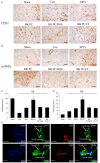
Effects of transplantation of BK-PC hEPCs on capillary and arteriole densities. Representative photographs of immunostaining by using CD31 (A) to identify capillaries and α-SMA (B) to identify arterioles are shown. (A: CD31, original magnification is 400 ×, bar for graph = 20 μm; B: α-SMA, original magnification is 200 ×, bar for graph = 50 μm.) Quantitative analysis of capillary density (C) and arteriole density (D) in the peri-infarct myocardium is also shown. All values are expressed as mean ± SEM (n = 5 for each group, *P < 0.01 versus other myocardial infarction groups). High-power field 2D (E) and 3D images (F) of DiD-labeled implanted hEPCs (red) and immunofluorescence staining of CD31 or α-SMA (green) at the border zone of the ischemic myocardium nuclei were counterstained with DAPI (blue). (Original magnification is 1000 ×; bar for graph = 20 μm).
Figure 4
Figure 4
Effects of BK-PC-hEPC transplantation on Akt, eNOS, VEGF expression, as well as NO production. A and B. Western blots analysis to identify Akt and eNOS expressions. C. NO production in myocardial tissue. D. Western blot analysis for VEGF expression. Representative blots are shown in the upper panel, and densitometric quantitation of protein expression levels are shown as fold changes in the lower panel. All values are expressed as mean ± SEM (n = 5 for each group; *P < 0.05 versus other myocardial infarction groups; #P < 0.01 versus other myocardial infarction groups).
Figure 5
Figure 5
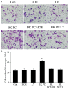
Effect of BK preconditioning on the migration of hEPCs. A. Representative photographs of the migrating hEPCs in the presence of 100 ng/mL SDF-1 stimulation among the six groups (original magnification: 200 ×). B. Quantitative analyses of the migrating hEPCs. All values are expressed as mean ± SEM. (n = 5 for each group; *P < 0.01 versus other groups).
Figure 6
Figure 6
Effects of BK preconditioning on capillary tube formation and vascular endothelial growth factor (VEGF) levels in cultured hEPCs. A. Representative photographs of the tube formation of Matrigel-coated cultured hEPCs in various groups (original magnification: 100 ×). B. Quantitative analysis of the mean tube length, which was measured by using Image-Pro Plus and was calculated against control groups. C. Enzyme-linked immunosorbent assay of VEGF levels in culture media. All values are expressed as mean ± SEM. (n = 5 for each group; *P < 0.01 versus other groups).

Similar articles

Cited by

References

    1. Ferrara N, Kerbel RS. Angiogenesis as a therapeutic target. Nature. 2005;438:967–974. - PubMed
    1. Annex BH. Therapeutic angiogenesis for critical limb ischaemia. Nat Rev Cardiol. 2013;10:387–396. - PubMed
    1. Sun YY, Bai WW, Wang B, Lu XT, Xing YF, Cheng W, Liu XQ, Zhao YX. Period 2 is essential to maintain early endothelial progenitor cell function in vitro and angiogenesis after myocardial infarction in mice. J Cell Mol Med. 2014;18:907–918. - PMC - PubMed
    1. Taljaard M, Ward MR, Kutryk MJ, Courtman DW, Camack NJ, Goodman SG, Parker TG, Dick AJ, Galipeau J, Stewart DJ. Rationale and design of Enhanced Angiogenic Cell Therapy in Acute Myocardial Infarction (ENACT-AMI): the first randomized placebo-controlled trial of enhanced progenitor cell therapy for acute myocardial infarction. Am Heart J. 2010;159:354–360. - PubMed
    1. Yu P, Li Q, Liu Y, Zhang J, Seldeen K, Pang M. Pro-angiogenic efficacy of transplanting endothelial progenitor cells for treating hindlimb ischemia in hyperglycemic rabbits. J Diabetes Complications. 2015;29:13–19. - PubMed

LinkOut - more resources