

Monosomy of chromosome 17 in breast cancer during interpretation of HER2 gene amplification
- PMID: 26328251
- PMCID: PMC4548332
Monosomy of chromosome 17 in breast cancer during interpretation of HER2 gene amplification
Abstract
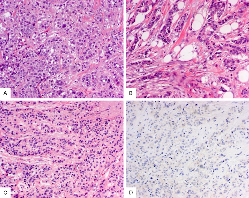
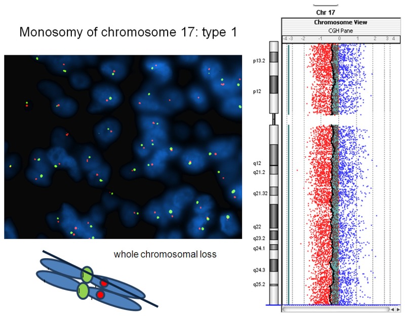
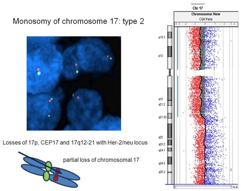
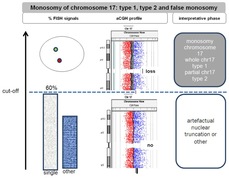
Monosomy of chromosome 17 may affect the assessment of HER2 amplification. Notably, the prevalence ranges from 1% up to 49% due to lack of consensus in recognition. We sought to investigate the impact of monosomy of chromosome 17 to interpretation of HER2 gene status. 201 breast carcinoma were reviewed for HER2 gene amplification and chromosome 17 status. FISH analysis was performed by using double probes (LSI/CEP). Absolute gene copy number was also scored per each probe. HER2 FISH test was repeated on serial tissue sections, ranging in thickness from 3 to 20 µm. Ratio was scored and subsequently corrected by monosomy after gold control test using the aCGH method to overcome false interpretation due to artefactual nuclear truncation. HER2 immunotests was performed on all cases. 26/201 cases were amplified (13%). Single signals per CEP17 were revealed in 7/201 (3.5%) cases. Five out of 7 cases appeared monosomic with aCGH (overall, 5/201, 2.5%) and evidenced single signals in >60% of nuclei after second-look on FISH when matching both techniques. Among 5, one case showed amplification with a pattern 7/1 (HER2/CEP17>2) of copies (3+ at immunotest); three cases revealed single signals per both probes (LSI/CEP=1) and one case revealed a 3:1 ratio; all last 4 cases showed 0/1+ immunoscore. We concluded that: 1) monosomy of chromosome 17 may be observed in 2.5% of breast carcinoma; 2) monosomy of chromosome 17 due to biological reasons rather than nuclear truncation was observed when using the cut-off of 60% of nuclei harboring single signals; 3) the skewing of the ratio due to single centromeric 17 probe may lead to false positive evaluation; 4) breast carcinomas showing a 3:1 ratio (HER2/CEP17) usually show negative 0/1+ immunoscore and <6 gene copy number at FISH.
Keywords: Breast carcinoma; FISH analysis; HER 2 amplification; aCGH method; chromosome 17; double probes; false positive; monosomy; ratio (HER2/CEP 17); single signals.
Figures
References
-
- Risio M, Casorzo L, Redana S, Montemurro F. HER2 gene-amplified breast cancers with monosomy of chromosome 17 are poorly responsive to trastuzumab-based treatment. Oncol Rep. 2005;13:305–9. - PubMed
-
- Marchio C, Lambros MB, Gugliotta P, Di Cantogno LV, Botta C, Pasini B, Tan DS, Mackay A, Fenwick K, Tamber N, Bussolati G, Ashworth A, Reis-Filho JS, Sapino A. Does chromosome 17 centromere copy number predict polysomy in breast cancer? A fluorescence in situ hybridization and microarray-based CGH analysis. J Pathol. 2009;219:16–24. - PubMed
-
- Starczynski J, Atkey N, Connelly Y, O’Grady T, Campbell FM, di Palma S, Wencyk P, Jasani B, Gandy M, Bartlett JM. HER2 gene amplification in breast cancer: a rogues’ gallery of challenging diagnostic cases: UKNEQAS interpretation guidelines and research recommendations. Am J Clin Pathol. 2012;137:595–605. - PubMed
-
- Wolff AC, Hammond ME, Hicks DG, Dowsett M, McShane LM, Allison KH, Allred DC, Bartlett JM, Bilous M, Fitzgibbons P, Hanna W, Jenkins RB, Mangu PB, Paik S, Perez EA, Press MF, Spears PA, Vance GH, Viale G, Hayes DF. Recommendations for human epidermal growth factor receptor 2 testing in breast cancer: American Society of Clinical Oncology/College of American Pathologists clinical practice guideline update. J. Clin. Oncol. 2013;31:3997–4013. - PubMed
LinkOut - more resources
Full Text Sources
Research Materials
Miscellaneous