Review
. 2016 Mar;36(2):155-65.
doi: 10.1007/s10571-015-0256-9. Epub 2015 Sep 2.

Endothelial Dysfunction and Amyloid-β-Induced Neurovascular Alterations

Affiliations
Review

Endothelial Dysfunction and Amyloid-β-Induced Neurovascular Alterations

Kenzo Koizumi et al. Cell Mol Neurobiol. 2016 Mar.

Abstract

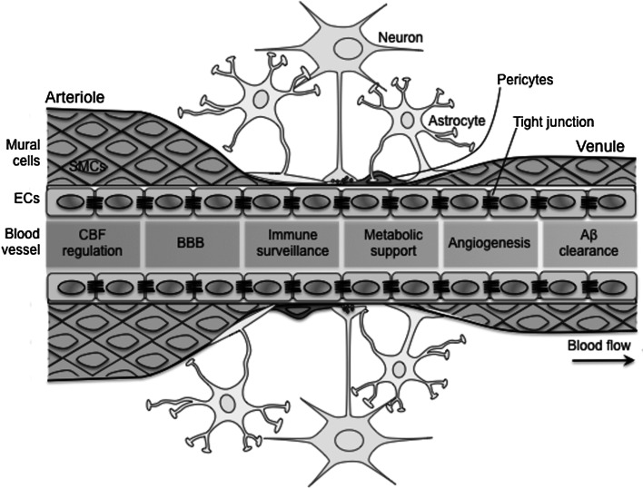
Alzheimer's disease (AD) and cerebrovascular diseases share common vascular risk factors that have disastrous effects on cerebrovascular regulation. Endothelial cells, lining inner walls of cerebral blood vessels, form a dynamic interface between the blood and the brain and are critical for the maintenance of neurovascular homeostasis. Accordingly, injury in endothelial cells is regarded as one of the earliest symptoms of impaired vasoregulatory mechanisms. Extracellular buildup of amyloid-β (Aβ) is a central pathogenic factor in AD. Aβ exerts potent detrimental effects on cerebral blood vessels and impairs endothelial structure and function. Recent evidence implicates vascular oxidative stress and activation of the non-selective cationic channel transient receptor potential melastatin (TRPM)-2 on endothelial cells in the mechanisms of Aβ-induced neurovascular dysfunction. Thus, Aβ triggers opening of TRPM2 channels in endothelial cells leading to intracellular Ca(2+) overload and vasomotor dysfunction. The cerebrovascular dysfunction may contribute to AD pathogenesis by reducing the cerebral blood supply, leading to increased susceptibility to vascular insufficiency, and by promoting Aβ accumulation. The recent realization that vascular factors contribute to AD pathobiology suggests new targets for the prevention and treatment of this devastating disease.

Keywords: Alzheimer’s disease; Cerebral blood flow; Cerebral endothelial cells; TRPM2; β-amyloid.


Figures

Fig. 1
Fig. 1
The neurovascular unit is formed of endothelial cells, mural cells (smooth muscle cells and pericytes), neurons, astrocytes, and others. The major function of neurovascular unit is to keep the brain in a homeostatic microenvironment. In arteries and capillaries, the astrocytes wrap the abluminal side of blood vessels with their foot processes. In capillaries, mural cells are replaced by pericytes. Endothelial cells line an inner layer of entire blood vessels and play a wide range of critical roles of vascular function in harmony with other neurovascular units and, thus, participate in all aspects of neurovascular homeostasis, including CBF regulation, BBB formation, immune surveillance, metabolic support, angiogenesis, and Aβ clearance. Aβ and cardiovascular risk factors damage the endothelial cells leading to the neurovascular, synaptic, and brain dysfunction. β-amyloid, BBB blood–brain barrier, ECs endothelial cells, SMCs smooth muscle cells
Fig. 2
Fig. 2
1-40 triggers large and sustained increases in intracellular Ca2+ via TRPM2 channels in brain endothelial cell. The Aβ1-40 (Aβ)-induced increases in intracellular Ca2+ are attenuated by the mechanistically distinct TRPM2 inhibitors 2-APB and ACA (a, b) or TRPM2 knockdown using siRNA, but not control siRNA (control si) (c, d). Data are presented as mean ± SEM. *p < 0.05; analysis of variance and Turkey’s test; N = 6–10/group. Modified from Park et al. (2014) with permission
Fig. 3
Fig. 3
Presumed mechanisms through which Aβ1-40 activates endothelial TRPM2 channels. Aβ1-40 (Aβ) activates the innate immunity receptor CD36 leading to production of superoxide via NADPH oxidase. Superoxide reacts with NO, made continuously in endothelial cells, to form peroxynitrite (PN). PN induces DNA damage, which, in turn, activates PARP. ADPR formation by PARG cleavage of PAR activates the Nudix (Nu) domain of TRPM2 leading to massive increases in intracellular Ca2+, which induce endothelial dysfunction. Modified from Park et al. (2014) with permission

Similar articles

Cited by

References

    1. Andresen J, Shafi NI, Bryan RM Jr (2006) Endothelial influences on cerebrovascular tone. J Appl Physiol 100(1):318–327. doi:10.1152/japplphysiol.00937.2005 - PubMed
    1. Ashe KH, Zahs KR (2010) Probing the biology of Alzheimer’s disease in mice. Neuron 66(5):631–645. doi:10.1016/j.neuron.2010.04.031 - PMC - PubMed
    1. Attwell D, Buchan AM, Charpak S, Lauritzen M, Macvicar BA, Newman EA (2010) Glial and neuronal control of brain blood flow. Nature 468(7321):232–243. doi:10.1038/nature09613 - PMC - PubMed
    1. Bateman GA, Levi CR, Schofield P, Wang Y, Lovett EC (2006) Quantitative measurement of cerebral haemodynamics in early vascular dementia and Alzheimer’s disease. J Clin Neurosci 13(5):563–568. doi:10.1016/j.jocn.2005.04.017 - PubMed
    1. Beach TG, Wilson JR, Sue LI, Newell A, Poston M, Cisneros R, Pandya Y, Esh C, Connor DJ, Sabbagh M, Walker DG, Roher AE (2007) Circle of Willis atherosclerosis: association with Alzheimer’s disease, neuritic plaques and neurofibrillary tangles. Acta Neuropathol 113(1):13–21. doi:10.1007/s00401-006-0136-y - PubMed

Substances

LinkOut - more resources