

Review
doi: 10.1152/physiol.00014.2015.
Motor System Reorganization After Stroke: Stimulating and Training Toward Perfection
Affiliations
- PMID: 26328881
- PMCID: PMC4556825
- DOI: 10.1152/physiol.00014.2015
Item in Clipboard
Review
Motor System Reorganization After Stroke: Stimulating and Training Toward Perfection
Physiology (Bethesda).
2015 Sep.
Abstract
Stroke instigates regenerative responses that reorganize connectivity patterns among surviving neurons. The new connectivity patterns can be suboptimal for behavioral function. This review summarizes current knowledge on post-stroke motor system reorganization and emerging strategies for shaping it with manipulations of behavior and cortical activity to improve functional outcome.
©2015 Int. Union Physiol. Sci./Am. Physiol. Soc.
Conflict of interest statement
No conflicts of interest, financial or otherwise, are declared by the author(s).
Figures
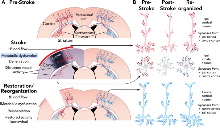
Mechanisms of spontaneous functional improvements after stroke A: coronal section illustrations of some of the axonal projections of cortical pyramidal neurons. Death of neurons in the core of an ischemic infarct results in denervation and disrupted activity in afferent targets, the striatum and contralateral cortex in this example. In peri-infarct cortex, there is a gradient of blood flow reductions, denervation of intracortical connections (not shown), and various degrees of dendritic retraction in surrounding neurons. Over time, blood flow and metabolic activity are restored, and denervated regions are reinnervated by axonal sprouting and synaptogenesis, resulting in reorganized connectivity. Restoration and reorganization are interrelated, e.g., reinnervation may depend on some degree of blood flow recovery and contribute to its fuller restoration. B: illustration of potential changes in neural connectivity patterns after reinnervation. The relative quantities of synapses from the contralateral vs. ipsilateral cortex may substantially change. Other sources of synaptic input to these neuronal populations can contribute to reinnervation and alter the balance of excitatory and inhibitory activity.
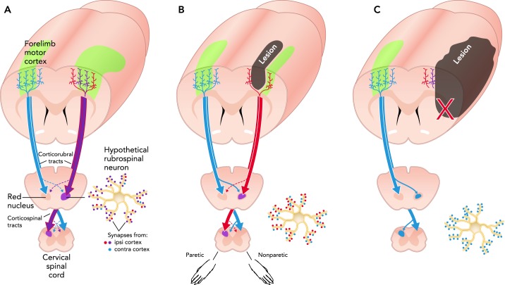
Neuroantomical reorganization after stroke A: illustration of a rat brain sectioned coronally near the rostral edge of the MI forelimb representation region (green). Callosal, corticorubral, and corticospinal tract projections of forelimb MI are illustrated in an intact brain. B: after subtotal infarcts of the forelimb area, remaining neurons of the forelimb region and surrounding motor cortex of the injured hemisphere can contribute to reinnervation. C: larger infarcts can severely damage descending projection pathways or the cortical pyramidal neurons that give rise to them, leaving crossed collaterals of contralesional projections as a primary source of reinnervation. The rubrospinal neuron illustration shows potential alterations in relative quantities of synaptic input from ipsi- and contralesional M1.
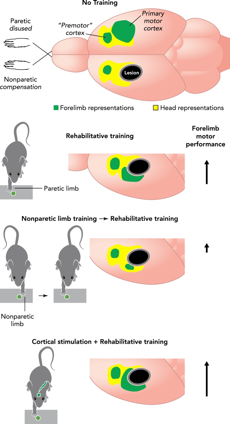
Skill training and cortical stimulation effects on motor maps of the paretic limb Motor skill training focused on the paretic forelimb (“rehabilitative training”) promotes the maintenance and restoration of forelimb movement representations, as well as the growth of dendrites and maturation of synapses in motor cortex. The effects of rehabilitative training are disrupted by prior training of the nonparetic limb (to model learning to compensate with this limb). The effects of rehabilitative training are amplified when electrical stimulation is delivered to cortex concurrently with training. Arrows indicate the magnitude of improvement in skilled motor function relative to no training.
References
-
- Adkins DL, Bury SD, Jones TA. Laminar-dependent dendritic spine alterations in the motor cortex of adult rats following callosal transection and forced forelimb use. Neurobiol Learn Mem 78: 35–52, 2002. - PubMed
-
- Adkins DL, Boychuk J, Remple MS, Kleim JA. Motor training induces experience-specific patterns of plasticity across motor cortex and spinal cord. J Appl Physiol 101: 1776–1782, 2006. - PubMed
-
- Adkins DL, Campos P, Quach D, Borromeo M, Schallert K, Jones TA. Epidural cortical stimulation enhances motor function after sensorimotor cortical infarcts in rats. Exp Neurol 200: 356–370, 2006. - PubMed
Publication types
MeSH terms
Grants and funding
LinkOut - more resources
Full Text Sources
Other Literature Sources
Medical