. 2016 Jan;69(1):15-25.
doi: 10.1038/ja.2015.77. Epub 2015 Sep 2.

Conserved biosynthetic pathways for phosalacine, bialaphos and newly discovered phosphonic acid natural products

Affiliations

Conserved biosynthetic pathways for phosalacine, bialaphos and newly discovered phosphonic acid natural products

Joshua A V Blodgett et al. J Antibiot (Tokyo). 2016 Jan.

Abstract

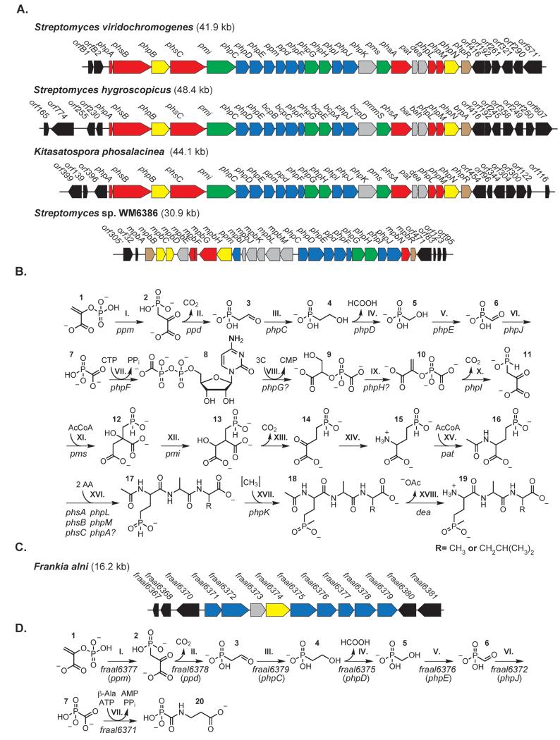
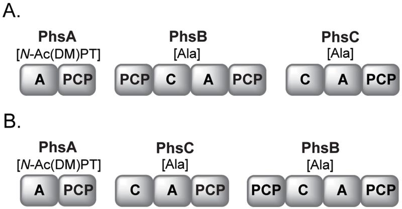
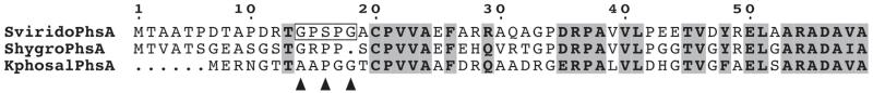
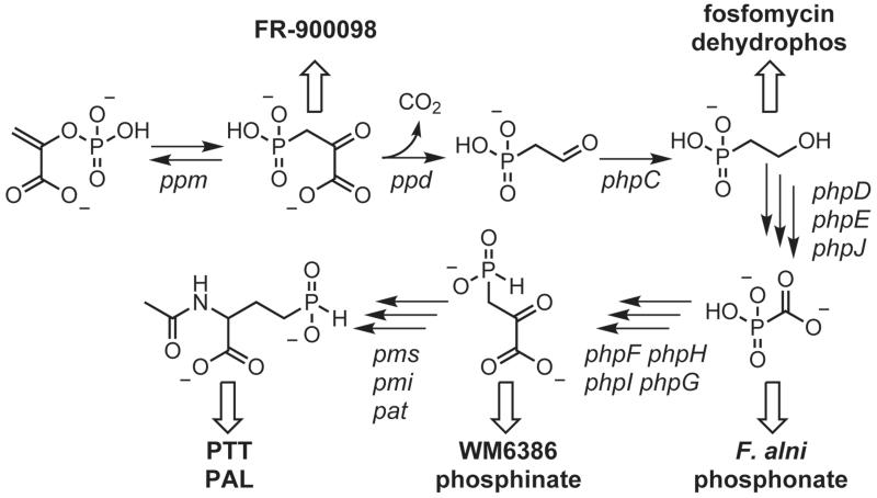
Natural products containing phosphonic or phosphinic acid functionalities often display potent biological activities with applications in medicine and agriculture. The herbicide phosphinothricin-tripeptide (PTT) was the first phosphinate natural product discovered, yet despite numerous studies, questions remain surrounding key transformations required for its biosynthesis. In particular, the enzymology required to convert phosphonoformate to carboxyphosphonoenolpyruvate and the mechanisms underlying phosphorus methylation remain poorly understood. In addition, the model for non-ribosomal peptide synthetase assembly of the intact tripeptide product has undergone numerous revisions that have yet to be experimentally tested. To further investigate the biosynthesis of this unusual natural product, we completely sequenced the PTT biosynthetic locus from Streptomyces hygroscopicus and compared it with the orthologous cluster from Streptomyces viridochromogenes. We also sequenced and analyzed the closely related phosalacine (PAL) biosynthetic locus from Kitasatospora phosalacinea. Using data drawn from the comparative analysis of the PTT and PAL pathways, we also evaluate three related recently discovered phosphonate biosynthetic loci from Streptomyces sviceus, Streptomyces sp. WM6386 and Frankia alni. Our observations address long-standing biosynthetic questions related to PTT and PAL production and suggest that additional members of this pharmacologically important class await discovery.


Figures

Figure 1
Figure 1. Comparison of phosphonate and phosphinate gene clusters and associated biosynthetic pathways
(A) Comparison of gene clusters encoding PTT and PAL biosynthetic enzymes with the phosphinate biosynthetic cluster of unknown function from Streptomyces sp. WM6386. Open reading frames shown in black do not have proposed roles in the biosynthesis of PTT or PAL. Most ORFs shown in blue have roles the synthesis and activation of phosphonoformate, an early intermediate. Also included is phpI (bcpA), which shares sequence and functional similarity to ppm, the gene associated with step I. Green ORFs are similar to those encoding enzymes utilized in glycolysis and the TCA cycle. Red ORFs encode proteins likely involved in non-ribosomal peptide synthesis. Yellow ORFs contain transmembrane domains. Grey ORFs likely encode proteins involved in tailoring reactions or self-resistance. ORFs encoding transcriptional regulators are colored brown. (B) Model for PTT and PAL biosyntheses adapted from multiple references and this work. ORF assignments to biosynthetic steps are based upon published work or by inference from gene similarity. Gene nomenclature from the S. viridochromogenes cluster is used throughout. Cofactors and water have been omitted for clarity. Label 3C in step VIII denotes the input of a three-carbon compound such as glycerate or 3-phosphoglycerate. Label “AA” in step XVI indicates the addition of two amino acid residues. For PTT biosynthesis, two alanine residues are added; for PAL one alanine and leucine each are incorporated. The brackets surrounding the methyl group in step XVII indicates the donor for this reaction has not rigorously been shown. (C) Putative phosphonate biosynthetic gene cluster from F. alni. (D) Hypothetical biosynthetic pathway for a predicted phosphonic acid metabolite deduced from F. alni genome mining. The GenBank locus identifiers are those from the published genome sequence. Homologs from early PTT biosynthesis allowing for plausible pathway prediction are noted in brackets.
Figure 2
Figure 2. Thiotemplate assembly and chain release models for PTT and PAL biosyntheses
Illustration showing NRPS domain architecture common to the PhsA, PhsB and PhsC enzymes of S. viridochromogenes, S. hygroscopicus and K. phosalacinea. PhsA from S. viridochromogenes loads both N-acetyldemethylphosphinothricin and N-acetylphosphinothricin; the desmethyl form is shown. The illustrated PhsA/B/C enzyme order is supported by the analyses in this work. PAL PhsC specificity for leucine awaits biochemical confirmation. Scheme A shows the use of either or possibly both PhpL and PhpM homologs in assembly line product release. Alternatively, one thioesterase homolog might be involved in product release where the other is involved in an editing role, as previously suggested. Scheme B shows a possible role for PTT/PAL PhpL homologs as PCP-to-PCP domain transacylases. An increasing number of Type II thioesterase homologs containing the variant GXCXG thioesterase motifs have been identified in natural product biosynthetic clusters where they function as transacylases. This model would then leave PhpM homologs containing typical GXSXG thioesterase motifs as product-releasing thioesterases. We note spontaneous hydrolysis might also contribute to chain release in both models.
Figure 3
Figure 3. Thioesterase signatures extracted from PTT and PAL biosynthetic genes
A)-Alignment of the first 59 N-terminal residues of PhsA from S. viridochromogenes, S. hygroscopicus and K. phosalacinea. The GXSXG motif hypothesized to be part of a mini-thioesterase domain in S. viridochromogenes PhsA is boxed. Arrowheads indicate equivalent positions in PhsAs from S.hygroscopicus and K. phosalacinea; the GXSXG motif found in the S. viridochromogenes synthetase is not conserved in the other PhsAs. Highly similar or identical residues are highlighted in grey. B) The alignment of catalytic motifs extracted from PTT/PAL PhpL homologs with those from Type II thioesterase-like transacylases involved in zorbamycin (ZbmVIId), syringomycin (SyrC), coronamic acid (CmaE) and bactobolin (BtaH) biosyntheses. Each contains a cysteine-centered GXCXG variant thioesterase motif (boxed, with consensus residues indicated by arrowheads). Residues surrounding the conserved catalytic histidine found in most Type II thioesterases (SviridoPhpL His223) are also shown.
Figure 4
Figure 4. Metabolic branchpoints from studied reduced phosphorus antibiotic biosyntheses
A number of phosphonate and phosphinate biosynthetic pathways share conserved early biosynthetic steps. Illustrated are a number of transformations common to multiple reduced phosphorus antibiotics and how they relate to the F. alni, WM6386 and PTT/PAL biosyntheses discussed in the text.

Similar articles

Cited by

References

    1. Metcalf WW, van der Donk WA. Biosynthesis of phosphonic and phosphinic acid natural products. Annu Rev Biochem. 2009;78:65–94. - PMC - PubMed
    1. De Clercq E. The Holy Trinity: the acyclic nucleoside phosphonates. Adv Pharmacol. 2013;67:293–316. - PubMed
    1. Kim SY, Ju KS, Metcalf WW, Evans BS, Kuzuyama T, van der Donk WA. Different biosynthetic pathways to fosfomycin in Pseudomonas syringae and Streptomyces species. Antimicrob Agents Chemother. 2012;56(8):4175–4183. - PMC - PubMed
    1. Eliot AC, et al. Cloning, expression, and biochemical characterization of Streptomyces rubellomurinus genes required for biosynthesis of antimalarial compound FR900098. Chem Biol. 2008;15(8):765–770. - PMC - PubMed
    1. Borisova SA, Circello BT, Zhang JK, van der Donk WA, Metcalf WW. Biosynthesis of rhizocticins, antifungal phosphonate oligopeptides produced by Bacillus subtilis ATCC6633. Chem Biol. 2010;17(1):28–37. - PMC - PubMed

Publication types

MeSH terms