

A dual-view digital tomosynthesis imaging technique for improved chest imaging
- PMID: 26328973
- PMCID: PMC4537485
- DOI: 10.1118/1.4928214
A dual-view digital tomosynthesis imaging technique for improved chest imaging
Abstract
Purpose: Digital tomosynthesis (DTS) has been shown to be useful for reducing the overlapping of abnormalities with anatomical structures at various depth levels along the posterior-anterior (PA) direction in chest radiography. However, DTS provides crude three-dimensional (3D) images that have poor resolution in the lateral view and can only be displayed with reasonable quality in the PA view. Furthermore, the spillover of high-contrast objects from off-fulcrum planes generates artifacts that may impede the diagnostic use of the DTS images. In this paper, the authors describe and demonstrate the use of a dual-view DTS technique to improve the accuracy of the reconstructed volume image data for more accurate rendition of the anatomy and slice images with improved resolution and reduced artifacts, thus allowing the 3D image data to be viewed in views other than the PA view.
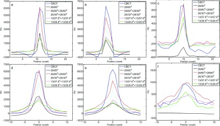
Methods: With the dual-view DTS technique, limited angle scans are performed and projection images are acquired in two orthogonal views: PA and lateral. The dual-view projection data are used together to reconstruct 3D images using the maximum likelihood expectation maximization iterative algorithm. In this study, projection images were simulated or experimentally acquired over 360° using the scanning geometry for cone beam computed tomography (CBCT). While all projections were used to reconstruct CBCT images, selected projections were extracted and used to reconstruct single- and dual-view DTS images for comparison with the CBCT images. For realistic demonstration and comparison, a digital chest phantom derived from clinical CT images was used for the simulation study. An anthropomorphic chest phantom was imaged for the experimental study. The resultant dual-view DTS images were visually compared with the single-view DTS images and CBCT images for the presence of image artifacts and accuracy of CT numbers and anatomy and quantitatively compared with root-mean-square-deviation (RMSD) values computed using the digital chest phantom or the CBCT images as the reference in the simulation and experimental study, respectively. High-contrast wires with vertical, oblique, and horizontal orientations in a PA view plane were also imaged to investigate the spatial resolutions and how the wire signals spread in the PA view and lateral view slice images.
Results: Both the digital phantom images (simulated) and the anthropomorphic phantom images (experimentally generated) demonstrated that the dual-view DTS technique resulted in improved spatial resolution in the depth (PA) direction, more accurate representation of the anatomy, and significantly reduced artifacts. The RMSD values corroborate well with visual observations with substantially lower RMSD values measured for the dual-view DTS images as compared to those measured for the single-view DTS images. The imaging experiment with the high-contrast wires shows that while the vertical and oblique wires could be resolved in the lateral view in both single- and dual-view DTS images, the horizontal wire could only be resolved in the dual-view DTS images. This indicates that with single-view DTS, the wire signals spread liberally to off-fulcrum planes and generated wire shadow there.
Conclusions: The authors have demonstrated both visually and quantitatively that the dual-view DTS technique can be used to achieve more accurate rendition of the anatomy and to obtain slice images with improved resolution and reduced artifacts as compared to the single-view DTS technique, thus allowing the 3D image data to be viewed in views other than the PA view. These advantages could make the dual-view DTS technique useful in situations where better separation of the objects-of-interest from the off-fulcrum structures or more accurate 3D rendition of the anatomy are required while a regular CT examination is undesirable due to radiation dose considerations.
Figures


References
Publication types
MeSH terms
Grants and funding
LinkOut - more resources
Full Text Sources
Other Literature Sources
