

Assessment of blood-brain barrier penetration of miltefosine used to treat a fatal case of granulomatous amebic encephalitis possibly caused by an unusual Balamuthia mandrillaris strain
- PMID: 26329128
- PMCID: PMC4676568
- DOI: 10.1007/s00436-015-4684-8
Assessment of blood-brain barrier penetration of miltefosine used to treat a fatal case of granulomatous amebic encephalitis possibly caused by an unusual Balamuthia mandrillaris strain
Abstract
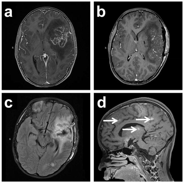
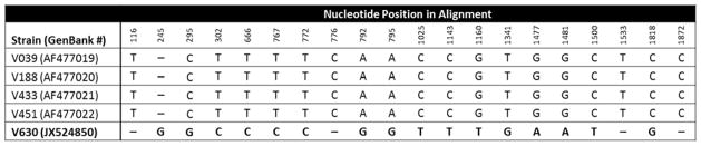
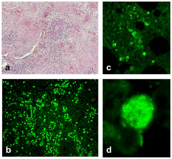
Balamuthia mandrillaris, a free-living ameba, causes rare but frequently fatal granulomatous amebic encephalitis (GAE). Few patients have survived after receiving experimental drug combinations, with or without brain lesion excisions. Some GAE survivors have been treated with a multi-drug regimen including miltefosine, an investigational anti-leishmanial agent with in vitro amebacidal activity. Miltefosine dosing for GAE has been based on leishmaniasis dosing because no data exist in humans concerning its pharmacologic distribution in the central nervous system. We describe results of limited cerebrospinal fluid (CSF) and serum drug level testing performed during clinical management of a child with fatal GAE who was treated with a multiple drug regimen including miltefosine. Brain biopsy specimens, CSF, and sera were tested for B. mandrillaris using multiple techniques, including culture, real-time polymerase chain reaction, immunohistochemical techniques, and serology. CSF and serum miltefosine levels were determined using a liquid chromatography method coupled to tandem mass spectrometry. The CSF miltefosine concentration on hospital admission day 12 was 0.4 μg/mL. The serum miltefosine concentration on day 37, about 80 h post-miltefosine treatment, was 15.3 μg/mL. These are the first results confirming some blood-brain barrier penetration by miltefosine in a human, although with low-level CSF accumulation. Further evaluation of brain parenchyma penetration is required to determine optimal miltefosine dosing for Balamuthia GAE, balanced with the drug's toxicity profile. Additionally, the Balamuthia isolate was evaluated by real-time polymerase chain reaction (PCR), demonstrating genetic variability in 18S ribosomal RNA (18S rRNA) sequences and possibly signaling the first identification of multiple Balamuthia strains with varying pathogenicities.
Keywords: Balamuthia; Encephalitis; Granulomatous; Miltefosine.
Conflict of interest statement
The authors declare that they have no conflicts of interest.
Figures
References
-
- Booton GC, Carmichael JR, Visvesvara GS, Byers TJ, Fuerst P. Genotyping of Balamuthia mandrillaris based on nuclear 18S and mitochondrial 16S rRNA genes. Am J Trop Med Hyg. 2003a;68:65–69. - PubMed
Publication types
MeSH terms
Substances
Grants and funding
LinkOut - more resources
Full Text Sources
Other Literature Sources
Medical
Molecular Biology Databases
