. 2015 Sep 2:13:69.
doi: 10.1186/s12915-015-0177-6.

Genome-wide replication landscape of Candida glabrata

Affiliations

Genome-wide replication landscape of Candida glabrata

Stéphane Descorps-Declère et al. BMC Biol. .

Abstract

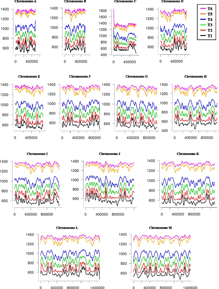
Background: The opportunistic pathogen Candida glabrata is a member of the Saccharomycetaceae yeasts. Like its close relative Saccharomyces cerevisiae, it underwent a whole-genome duplication followed by an extensive loss of genes. Its genome contains a large number of very long tandem repeats, called megasatellites. In order to determine the whole replication program of the C. glabrata genome and its general chromosomal organization, we used deep-sequencing and chromosome conformation capture experiments.

Results: We identified 253 replication fork origins, genome wide. Centromeres, HML and HMR loci, and most histone genes are replicated early, whereas natural chromosomal breakpoints are located in late-replicating regions. In addition, 275 autonomously replicating sequences (ARS) were identified during ARS-capture experiments, and their relative fitness was determined during growth competition. Analysis of ARSs allowed us to identify a 17-bp consensus, similar to the S. cerevisiae ARS consensus sequence but slightly more constrained. Megasatellites are not in close proximity to replication origins or termini. Using chromosome conformation capture, we also show that early origins tend to cluster whereas non-subtelomeric megasatellites do not cluster in the yeast nucleus.

Conclusions: Despite a shorter cell cycle, the C. glabrata replication program shares unexpected striking similarities to S. cerevisiae, in spite of their large evolutionary distance and the presence of highly repetitive large tandem repeats in C. glabrata. No correlation could be found between the replication program and megasatellites, suggesting that their formation and propagation might not be directly caused by replication fork initiation or termination.


Figures

Fig. 1
Fig. 1
Sequence coverage during S-phase time course. The sequence coverage at each time point (T1 to T6) for each of the 13 chromosomes, is shown. For each nucleotide (on the x axis) at every time point, relative sequence coverage is plotted (on the y axis) as the ratio of Tn coverage over T0 coverage (see “Methods”). Early replication origins are visible as peaks in the first time points, and gradually disappear when regions between origins are being replicated
Fig. 2
Fig. 2
Replication landscape of C. glabrata chromosomes. For each of the 13 chromosomes (labeled from a to m), the T50 of each nucleotide is shown as a black line, peaks correspond to replication origins, and valleys to termini. Post-elutriation times, after release in fresh medium, are indicated on the right side of each graph (in minutes). The GC content of each chromosome is drawn as a red line, with GC % indicated on the left side of each graph (5 kb sliding windows by 500 bp steps). The distance between two gray vertical bars under each graph is 250 kb. Dotted vertical lines represent centromere positions. MAT, HML, and HMR loci are shown as blue boxes, at their location as determined by Muller et al. [76]. Histone genes are shown as orange boxes. Subtelomeric locations of both rDNA tandem arrays are indicated by green arrows on chromosomes L and M. Autonomously replicating sequences positions are indicated above each graph as red dots
Fig. 3
Fig. 3
Replication timing of chromosomal features. a Distribution of early and late origins, according to their T50. The percentage of each class is represented on the y axis. Each interval corresponds to 30 s. Early and late origins are defined according to the observed distribution of the two populations of origins, origins firing before 69 min being labeled as early. The distribution of bona fide origins is shown in light orange, and is not statistically different from the whole distribution. b Determination of the average initial replication fork speed. Average fork rates, shown as black dots, are plotted every 400 bp according to their distance from early origins (only bona fide early origins were considered). The orange line corresponds to the linear regression of the first 2 kb. The red line corresponds to the linear regression of the first 5 kb. Correlation coefficients (r2) are indicated near each line. The blue line corresponds to the baseline at which no replication occurs. Intersection of each linear regression with the baseline indicates the amount of DNA replicated within a 5-min time frame (10.5 kb or 15.1 kb) for an average fork speed between 2.1 kb/min and 3 kb/min. c Distribution of replication timing of centromeres and telomeres, according to their T50. Intervals to which mating-type cassettes belong are indicated by blue arrows and histone genes by orange arrows. Note that “Early” and “Late” replication time frames correspond to those defined in a. d Distribution of replication timing of internal chromosomal deletions and chromosomal breakpoints found in translocations, according to their T50. e Distribution of replication timing of subtelomeric and internal megasatellites, according to their T50
Fig. 4
Fig. 4
Replication timing of chromosomal arms. a For each chromosome arm, T50 are represented as boxplots. Red: left chromosome arm values; blue: right chromosome arm values. b Phylogeny of C-left and right arm genes. Average Z-scores of distances between C. glabrata genes and closely related yeast species are shown for each chromosome C arm. None of these distances was significantly different from the other (t-test p-values of C left Z-scores Nc vs Kp, 0.93; Nc vs Zr, 0.79; t-test p-values of C right Z-scores Nc vs Kp, 0.90; Nc vs Zr, 0.70; significance threshold 0.05). Kp Kluyveromyces polysporus, Nc Naumovozyma castellii, Zr Zygosaccharomyces rouxii
Fig. 5
Fig. 5
ARS capture and fitnesses. a Distribution of distances between ARSs and replication origins. The observed distribution is shown in blue. The simulation of 1,000 independent experiments is shown in gray (see text). The dotted red line corresponds to the 3-kb distance limit that was chosen to define bona fide origins. b Coverage of each ARS at G50 (y axis) as compared to G0 (x axis). The dotted red line corresponds to a coverage ratio 1/1. ARSs with the highest G50 coverage are labeled. c Same as b, but for G100 coverage of each ARS (y axis), as compared to G50 coverage (x axis). ARS autonomously replicating sequence
Fig. 6
Fig. 6
ARS consensus sequences. a C. glabrata ACS determined from the whole set of 275 ARSs (top), or from the 83 bona fide origins (bottom). Boundaries of the element are clearly visible in the latter case. The three positions differing from the S. cerevisiae ACS are shown by orange arrows. The WTW trinucleotide was barely detected. b S. cerevisiae ACS determined from the set of 337 known ARSs listed in the Saccharomyces Genome Database (SGD) [70] (top), or from the 274 ARSs identified here (bottom). Boundaries of the canonical ACS [10] or of the extended ACS (EACS) are indicated, as well as the B1 box and WTW trinucleotide [38]. Note that although the ACS identified in the ARS capture experiment was closer to the C. glabrata ACS than to the S. cerevisiae canonical ACS, the requirement for the B1 box was greater in S. cerevisiae than in C. glabrata. c Alignment of ARS_F6 with the ARS identified by Zordan et al. [46], showing 48 bp in common between the two sequences. The ACS is shown in red. ACS ARS consensus sequence, ARS autonomously replicating sequence
Fig. 7
Fig. 7
Chromatin organization of C. glabrata exponentially growing cells. a Normalized genomic contact matrix obtained from an asynchronous growing population. The 13 chromosomes are indicated on the x axis. The color scale on the right indicates the frequency of interactions between two regions of the genome (blue rare contacts; white frequent contacts; red very frequent contacts; exponential scale). Red arrowheads centromere clustering. Yellow arrowheads telomere contacts. b Colocalization Score (CS) for DNA regions of interest. The CS is the mean of measured contacts between DNA regions. The boxplots represent the distribution of CS expected by chance, obtained with 1,000 randomized sets of positions (keeping overall chromosomal distribution), whereas the red dot corresponds to the CS of each group of interest (Megasatellites, ARSs, early and late origins). Statistical significance of colocalization is attained when the red dot lies above the random group distribution (p-value < 0.001). c Nucleosomal organization at bona fide origins. Nucleosome signals 800 bp upstream and downstream of bona fide origins were aligned (top), the color code representing nucleosome density (blue low density; red high density). The bottom curve represents the average value of nucleosome density. Regular nucleosome spacing is observed, with a large depletion at replication origins (red arrow). Note that ACS positions (instead of ARS/origins positions) were used, to increase resolution. d Nucleosomal organization at all ARSs. Nucleosome density around all ACS positions were determined and also show a significant depletion. ACS ARS consensus sequence, ARS autonomously replicating sequence

Similar articles

Cited by

References

    1. Chan CS, Tye BK. Autonomously replicating sequences in Saccharomyces cerevisiae. Proc Natl Acad Sci U S A. 1980;77:6329–33. doi: 10.1073/pnas.77.11.6329. - DOI - PMC - PubMed
    1. Brewer BJ, Fangman WL. The localization of replication origins on ARS plasmids in S. cerevisiae. Cell. 1987;51:463–71. doi: 10.1016/0092-8674(87)90642-8. - DOI - PubMed
    1. Poloumienko A, Dershowitz A, De J, Newlon CS. Completion of replication map of Saccharomyces cerevisiae chromosome III. Mol Biol Cell. 2001;12:3317–27. doi: 10.1091/mbc.12.11.3317. - DOI - PMC - PubMed
    1. Donato JJ, Chung SC, Tye BK. Genome-wide hierarchy of replication origin usage in Saccharomyces cerevisiae. PLoS Genet. 2006;2 doi: 10.1371/journal.pgen.0020141. - DOI - PMC - PubMed
    1. Liachko I, Tanaka E, Cox K, Chung SC, Yang L, Seher A, et al. Novel features of ARS selection in budding yeast Lachancea kluyveri. BMC Genomics. 2011;12:633. doi: 10.1186/1471-2164-12-633. - DOI - PMC - PubMed

LinkOut - more resources