. 2015;16 Suppl 11(Suppl 11):S8.
doi: 10.1186/1471-2105-16-S11-S8. Epub 2015 Aug 13.

Physically-based in silico light sheet microscopy for visualizing fluorescent brain models

Physically-based in silico light sheet microscopy for visualizing fluorescent brain models

Marwan Abdellah et al. BMC Bioinformatics. 2015.

Abstract

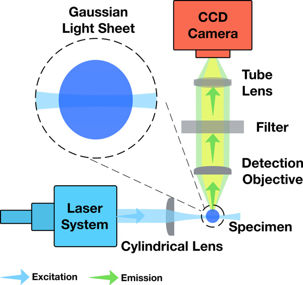
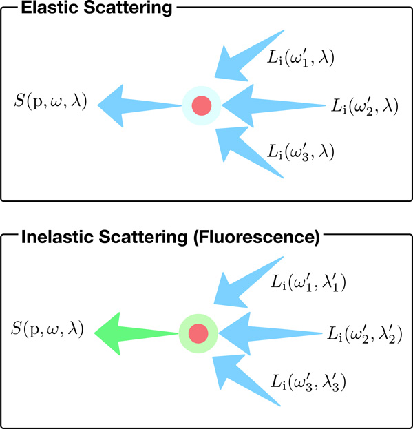
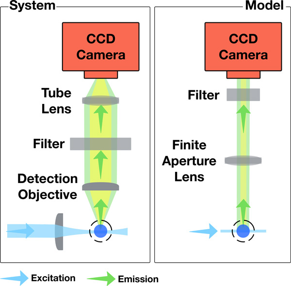
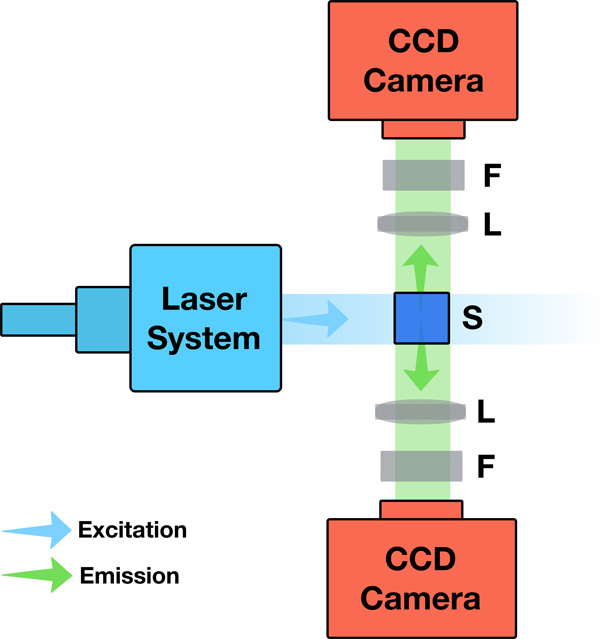
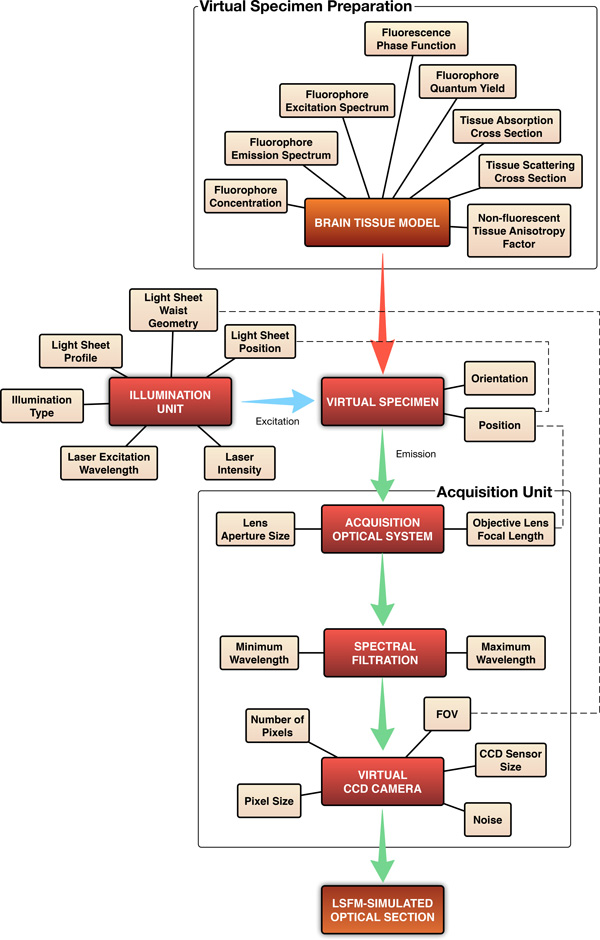
Background: We present a physically-based computational model of the light sheet fluorescence microscope (LSFM). Based on Monte Carlo ray tracing and geometric optics, our method simulates the operational aspects and image formation process of the LSFM. This simulated, in silico LSFM creates synthetic images of digital fluorescent specimens that can resemble those generated by a real LSFM, as opposed to established visualization methods producing visually-plausible images. We also propose an accurate fluorescence rendering model which takes into account the intrinsic characteristics of fluorescent dyes to simulate the light interaction with fluorescent biological specimen.

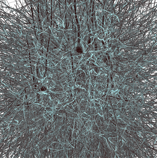
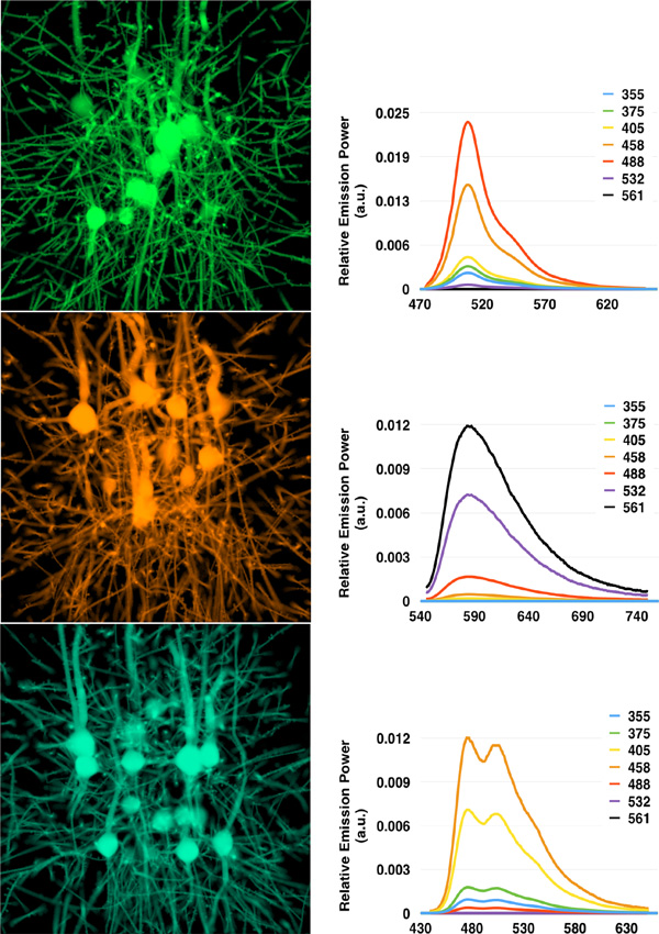
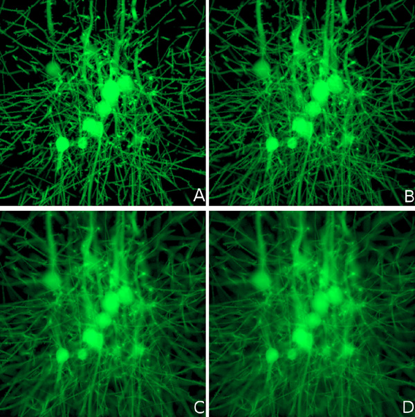
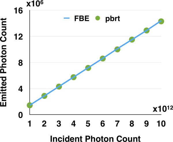
Results: We demonstrate first results of our visualization pipeline to a simplified brain tissue model reconstructed from the somatosensory cortex of a young rat. The modeling aspects of the LSFM units are qualitatively analysed, and the results of the fluorescence model were quantitatively validated against the fluorescence brightness equation and characteristic emission spectra of different fluorescent dyes.

Ams subject classification: Modelling and simulation.


Figures

Figure 1
Figure 1
A simplified top view diagram of the LSFM showing its main components and relative optical setup. The close up highlights the approximate rectangular profile of a Gaussian sheet intersection with the specimen.
Figure 2
Figure 2
Simple representation for elastic and inelastic scattering events at a point p.
Figure 3
Figure 3
Comparison between the acquisition system of the LSFM and our model. The coupling between the detection objective lens and the tube lens is modeled by an orthographic camera.
Figure 4
Figure 4
Surface rendering of a neuronal mesh model extracted from a microcircuit reconstructed from the cortex of a young rat. The size of this block is 100 µm3. The virtual specimen was created by converting this model into a fluorescent tagged-volume using solid voxelization.
Figure 5
Figure 5
In silico optical sectioning. (Left) Synthetic optical sections generated from the GFP- (top), RFP- (middle), and CFP-tagged (bottom) virtual specimens excited with maximum excitation wavelength. (Right) Emission SPDs measured from the rendered images at different excitation wavelengths between 355 and 561 nm. The curves are normalized to the SPD resulting at the maximum excitation wavelength for each respective case. The x-axis of the SPDs represents the wavelength in nm.
Figure 6
Figure 6
Rendering an optical section from the GFP-tagged specimen with multiple light sheet thicknesses: (A) 5, (B) 7.5, (C) 10, and (D) 12.5 µm. The increased blur with thicker light sheets is due to the detection of out-of-focus light rays.
Figure 7
Figure 7
The configuration and optical setup of the LSFM in the validation experiment. S, F and L indicate the specimen, the filter, and the lens with finite aperture.
Figure 8
Figure 8
Comparison between the number of photons calculated from the FBE and those detected from the simulation in PBRT using our fluorescence model.
Figure 9
Figure 9
An overview of the workflow of our visualization system and its parameters.

References

    1. Ahrens MB, Orger MB, Robson DN, Li JM, Keller PJ. Whole-brain functional imaging at cellular resolution using light-sheet microscopy. Nature Methods. 2012;10(5):413–420. doi: 10.1038/nmeth.2434. - DOI - PubMed
    1. Santi PA. Light sheet fluorescence microscopy: A review. Journal of Histochemistry & Cytochemistry. 2011;59(2):129–138. doi: 10.1369/0022155410394857. - DOI - PMC - PubMed
    1. Verveer PJ, Swoger J, Pampaloni F, Greger K, Marcello M, Stelzer EH. High-resolution three-dimensional imaging of large specimens with light sheet-based microscopy. Nature methods. 2007;4(4):311–313. - PubMed
    1. Santi PA, Johnson SB, Hillenbrand M, GrandPre PZ, Glass TJ, Leger JR. Thin-sheet laser imaging microscopy for optical sectioning of thick tissues. Biotechniques. 2009;46(4):287. - PMC - PubMed
    1. Tomer R, Ye L, Hsueh B, Deisseroth K. Advanced CLARITY for rapid and high-resolution imaging of intact tissues. Nat Protocols. 2014;9(7):1682–1697. doi: 10.1038/nprot.2014.123. - DOI - PMC - PubMed

Publication types

Substances