. 2015;88(1055):20150224.
doi: 10.1259/bjr.20150224. Epub 2015 Sep 2.

Evaluation of lung cancer by enhanced dual-energy CT: association between three-dimensional iodine concentration and tumour differentiation

Affiliations

Evaluation of lung cancer by enhanced dual-energy CT: association between three-dimensional iodine concentration and tumour differentiation

Shingo Iwano et al. Br J Radiol. 2015.

Abstract

Objective: To investigate the correlation between iodine concentration of dual-energy CT (DECT) and histopathology of surgically resected primary lung cancers.

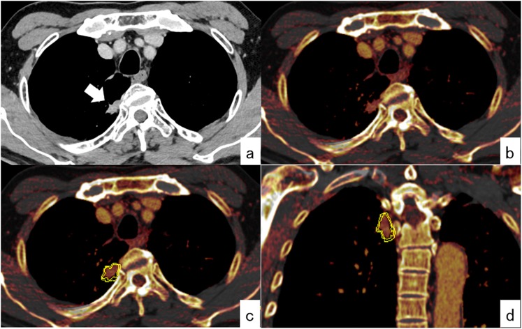
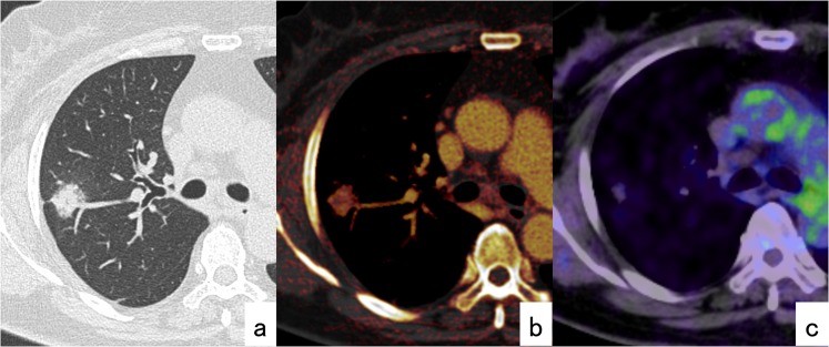
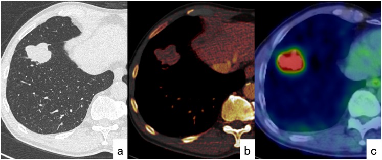
Methods: We reviewed the medical records, post-operative pathological records and pre-operative DECT images of patients who underwent surgical lung resection for primary lung cancer. After injection of iodinated contrast media, arterial and delayed phases were scanned using 140- and 80-kV tube voltages. Three-dimensional iodine concentration (iodine volume) of primary tumours was calculated using lung nodule application software.

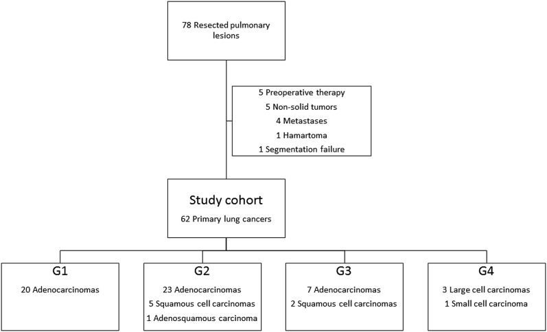
Results: A total of 60 patients (37 males and 23 females; age range, 39-84 years; mean age, 69 years) with 62 lung cancers were analysed. The resected tumours were histopathologically classified into well-differentiated (G1; n = 20), moderately differentiated (G2; n = 29), poorly differentiated (G3; n = 9) and undifferentiated (G4; n = 4) groups by degree of tumour differentiation (DTD). The mean ± standard deviation of iodine volume at the delayed phase was 59.6 ± 18.6 HU in G1 tumours, 46.5 ± 11.3 HU in G2 tumours, 34.3 ± 15.0 HU in G3 tumours and 28.8 ± 6.4 HU in G4 tumours; significant differences were observed between groups (p < 0.001). Univariate logistic regression analysis showed that iodine volumes both at the early and delayed phases were significantly correlated with DTD (p = 0.006 and p = 0.001, respectively), whereas gender, body weight and tumour size were not (p = 0.084, p = 0.062 and p = 0.391, respectively).

Conclusion: The iodine volume of lung cancers was significantly associated with their DTD. High-grade tumours tended to have lower iodine volumes than low-grade tumours.

Advances in knowledge: Iodine volume measured by DECT could be a valuable functional imaging method to estimate differentiation of primary lung cancer.


Figures

Figure 1.
Figure 1.
Procedure for measuring the iodine volume of lung lesions. (a) At the delayed phase of contrast-enhanced CT, a solid-type adenocarcinoma is observed in the right upper lobe (arrow). (b) Iodine-enhanced images at the same level as shown in (a). (c) Three-dimensional measurement of the iodine value on an iodine-enhanced image. Automatic segmentation (double yellow line) and calculation of iodine volume can be performed using the “Lung Nodule” application of dual-energy CT. (d) Coronal view of iodine-enhanced image after segmentation.
Figure 2.
Figure 2.
Study cohort flow chart. A total of 78 surgically resected pulmonary lesions were scanned pre-operatively with dual-phase contrast-enhanced CT using a dual-source technique. After exclusion, 62 primary lung cancers were included in the present analysis.
Figure 3.
Figure 3.
A 62-year-old female patient. (a) Well-differentiated (G1) adenocarcinoma is in the right upper lobe. (b) Iodine-enhanced image at the delayed phase of contrast-enhanced CT. The iodine volume is 57 HU. (c) Positron emission tomography/CT shows low fludeoxyglucose uptake of the tumour and the maximal standard uptake value is 1.1.
Figure 4.
Figure 4.
A 65-year-old male patient. (a) Poorly differentiated (G3) adenocarcinoma is in the right lower lobe. (b) Iodine-enhanced image at the delayed phase of contrast-enhanced CT. The iodine volume is 20 HU. (c) Positron emission tomography/CT shows high fludeoxyglucose uptake of the tumour and the maximal standard uptake value is 10.1.

Similar articles

Cited by

References

    1. Chung CK, Zaino R, Stryker JA, O'Neill M, Jr, DeMuth WE, Jr. Carcinoma of the lung: evaluation of histological grade and factors influencing prognosis. Ann Thorac Surg 1982; 33: 599–604. doi: 10.1016/S0003-4975(10)60819-3 - DOI - PubMed
    1. Kozu Y, Maniwa T, Takahashi S, Isaka M, Ohde Y, Nakajima T. Risk factors for both recurrence and survival in patients with pathological stage I non-small-cell lung cancer. Eur J Cardiothorac Surg 2013; 44: e53–8. doi: 10.1093/ejcts/ezt192 - DOI - PubMed
    1. Kishimoto M, Iwano S, Ito S, Kato K, Ito R, Naganawa S. Prognostic evaluations of small size lung cancers by 18F-FDG PET/CT and thin-section CT. Lung Cancer 2014; 86: 180–4. doi: 10.1016/j.lungcan.2014.09.006 - DOI - PubMed
    1. Lee HY, Jeong JY, Lee KS, Yi CA, Kim BT, Kang H, et al. . Histopathology of lung adenocarcinoma based on new IASLC/ATS/ERS classification: prognostic stratification with functional and metabolic imaging biomarkers. J Magn Reson Imaging 2013; 38: 905–13. doi: 10.1002/jmri.24080 - DOI - PubMed
    1. Grgic A, Yuksel Y, Groschel A, Schafers HJ, Sybrecht GW, Kirsch CM, et al. . Risk stratification of solitary pulmonary nodules by means of PET using (18)F-fluorodeoxyglucose and SUV quantification. Eur J Nucl Med Mol Imaging 2010; 37: 1087–94. doi: 10.1007/s00259-010-1387-3 - DOI - PubMed