Review
. 2015 Sep 1;142(17):2864-75.
doi: 10.1242/dev.119768.

Neuromesodermal progenitors and the making of the spinal cord

Affiliations
Review

Neuromesodermal progenitors and the making of the spinal cord

Domingos Henrique et al. Development. .

Abstract

Neuromesodermal progenitors (NMps) contribute to both the elongating spinal cord and the adjacent paraxial mesoderm. It has been assumed that these cells arise as a result of patterning of the anterior neural plate. However, as the molecular mechanisms that specify NMps in vivo are uncovered, and as protocols for generating these bipotent cells from mouse and human pluripotent stem cells in vitro are established, the emerging data suggest that this view needs to be revised. Here, we review the characteristics, regulation, in vitro derivation and in vivo induction of NMps. We propose that these cells arise within primitive streak-associated epiblast via a mechanism that is separable from that which establishes neural fate in the anterior epiblast. We thus argue for the existence of two distinct routes for making central nervous system progenitors.

Keywords: Bipotent cells; FGF; Neural induction; Neuromesodermal progenitors; Spinal cord; Stem cells; Wnt.


Conflict of interest statement

Competing interests

The authors declare no competing or financial interests.

Figures
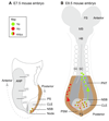
Fig. 1
Fig. 1. Key features of the developing CNS and neuromesodermal progenitors in the embryo.
Schematics of E7.5 (A) and E8.5 (B) mouse embryos indicating cell populations that give rise to the CNS. At E7.5, the anterior neural plate (ANP) consists of prospective forebrain (FB), midbrain (MB), hindbrain (HB) and some anterior spinal cord (aSC) progenitors; more posterior spinal cord arises from neuromesodermal progenitors (NMps; red/green), which are located in the node-streak border (NSB) in the anterior primitive streak (PS; brown) and in the adjacent caudal lateral epiblast (CLE; light grey). At E8.5, NMps have given rise to new neural progenitors (Np; green), which contribute to the CLE (light grey) and then the preneural tube (PNT; dark grey), and to new mesoderm progenitors (Mp; red), which contribute to presomitic mesoderm (PSM; brown). The rostralmost position reported for Nps generated by NMps is the ventral region of the anterior spinal cord approximately at the level of somite 6 (S6).
Fig. 2
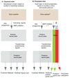
Fig. 2. Comparison of neural induction models.
(A) Prevailing view of vertebrate neural induction based on work in the amphibian embryo. This model, derived from Nieuwkoop's ‘activation-transformation’ hypothesis, involves the induction of an initial anterior neural plate that is subsequently regionalised by posteriorising signals to form posterior neural plate. (B) Proposed view of neural induction involving a dual origin of neural progenitors. In this model, epiblast cells (which in chick may have entered an unstable ‘preneural’ state, indicated by the asterisk) acquire neural fate either in the anterior neural plate (which is then progressively subdivided as proposed by Nieuwkoop) or via the induction of primitive streak-associated neuromesodermal progenitors (NMps), which contribute progenitors to anterior and posterior spinal cord and to flanking presomitic mesoderm (see text for details).
Fig. 3
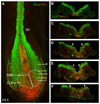
Fig. 3. Sox2 and brachyury co-expressing cells in the CLE and primitive streak.
(A) Confocal maximum intensity projection of the posterior end of an E8.5 (6-somite, S6) mouse embryo labelled with antibodies against Sox2 (green) and brachyury (Bra; red). Note the double-labelled cells in the CLE (white dashed lines) and NSB. (B-F) Transverse sections at the levels indicated in A. Note the double-labelled cells in the primitive streak and adjacent CLE (between the arrowheads). Sox2 is also detected in large, ventrally located migrating germ cells.
Fig. 4
Fig. 4. Key signals and transcriptional networks regulating NMps.
FGF and Wnt signals provided by the primitive streak and CLE induce the expression of Bra and the Sox2 (N1) enhancer, and Bra in turn promotes Wnt signalling. FGF signalling also promotes expression of Nkx1.2 (Sax1), and this transcription factor in turn induces Fgf8 transcription; it also indirectly promotes Wnt signalling by inhibiting expression of the repressor Tcf3 [indicated with a dotted line as evidence comes from P19 cells (Tamashiro et al., 2012)]. Wnt signalling induces the expression of Cdx genes, which act both to promote Wnt signalling and to regulate caudal Hox gene expression. Sox2 transcription is also repressed by BMP signalling delivered by epiblast cells posterior and lateral to the CLE and so defines the domain within which NMps can arise. The co-expression of Sox2 and Bra is a central feature of NMps and there is some evidence that they are mutually repressive (indicated by dotted inhibition symbols). For example, Sox2 mRNA expression is high in Bra mutant NMps in which Wnt is activated (Gouti et al., 2014); in the frog, T-box genes directly repress Sox2 (Gentsch et al., 2013); and in the mouse the presomitic mesoderm gene Tbx6 represses Sox2 via the N1 enhancer (Li and Storey, 2011; Takemoto et al., 2011). Conversely, Sox2 N1 loss (in a Sox3 null background) increases the ingression of cells to form presomitic mesoderm (Yoshida et al., 2014), suggesting that Sox2 normally restrains this Bra-induced activity; Sox2 also binds the Bra promoter in ESC-derived neural progenitors and Sox2 overexpression represses Bra in a Wnt-driven mesodermal differentiation assay (Zhao et al., 2004; Thomson et al., 2011). This mutual repression between Sox2 and Bra might underpin the creation of a state in which cells are poised to adopt either neural or mesodermal cell fate.
Fig. 5
Fig. 5. In vitro generation of NMps.
Summary of protocols used in recent studies to generate NMps in vitro from pluripotent mouse or human cells. The application of exogenous molecules over time is detailed, as well as the matrix used to plate the cells. The percentage of Bra/Sox2 co-expressing cells observed in the NMp population is also indicated. Blue bars, medium base; orange bars, FGF regime; red bars, the addition of CHIR99021 (a GSK3β inhibitor, used for Wnt signalling activation); purple bar, the addition of SB431542 [an inhibitor of the activin receptor-like kinase receptors ALK4/5/7 (Acvr1b/Tgfβr1/Acvr1c)]. EpiSC medium refers to a DMEM-based medium containing activin A and FGF2. Note that Tsakiridis et al. (2014) obtained NMps after either 48 h or 72 h incubation in the differentiation regime (asterisks). Lippmann et al. (2015) maintained theNMp regime (FGF2+CHIR99021) for up to 168 h (7 days), generating progenitors with progressively more posterior identities. All studies varied/optimised culture conditions for the organism/cell line used. For detailed information about the individual protocols (including concentrations of exogenous molecules applied), refer to the original publications. m, mouse; h, human; ESC, embryonic stem cell; EpiSC, epiblast-derived stem cells; PSC, pluripotent stem cell; nd, not determined.
Fig. 6
Fig. 6. Summary of events contributing to the acquisition of neural fate in the anterior epiblast and to NMp formation.
(A) Steps taking place in an E7.5 mouse embryo epiblast. The key steps leading to the acquisition of neural fate in the anterior neural plate (ANP; grey) and to NMp induction in the caudal lateral epiblast/node-streak border (CLE/NSB; light grey) are indicated. The primitive streak (PS) is also shown (brown). (B) The key gene regulatory networks (GRNs) predicted to be operating in each region, based on analyses in differentiating mouse EpiSCs (Iwafuchi-Doi et al., 2012).

References

    1. Akai J, Halley PA, Storey KG. FGF-dependent Notch signaling maintains the spinal cord stem zone. Genes Dev. 2005;19:2877–2887. - PMC - PubMed
    1. Anderson MJ, Naiche LA, Wilson CP, Elder C, Swing DA, Lewandoski M. TCreERT2, a transgenic mouse line for temporal control of Cre-mediated recombination in lineages emerging from the primitive streak or tail bud. PLoS ONE. 2013;8:e62479. - PMC - PubMed
    1. Andoniadou CL, Martinez-Barbera JP. Developmental mechanisms directing early anterior forebrain specification in vertebrates. Cell Mol Life Sci. 2013;70:3739–3752. - PMC - PubMed
    1. Ang SL, Conlon RA, Jin O, Rossant J. Positive and negative signals from mesoderm regulate the expression of mouse Otx2 in ectoderm explants. Development. 1994;120:2979–2989. - PubMed
    1. Aulehla A, Wehrle C, Brand-Saberi B, Kemler R, Gossler A, Kanzler BG, Herrmann BG. Wnt3a plays a major role in the segmentation clock controlling somitogenesis. Dev Cell. 2003;4:395–406. - PubMed

Publication types